 Login / Register


- Clone
- L243 (See other available formats)
- Regulatory Status
- RUO
- Other Names
- Major Histocompatibility Class II, MHC class II
- Isotype
- Mouse IgG2a, κ
- Ave. Rating
- Submit a Review
- Product Citations
- publications
HLA-DR is a heterodimeric cell surface glycoprotein comprised of a 36 kD α (heavy) chain and a 27 kD β (light) chain. It is expressed on B cells, activated T cells, monocytes/macrophages, dendritic cells, and other non-professional APCs. In conjunction with the CD3/TCR complex and CD4 molecules, HLA-DR is critical for efficient peptide presentation to CD4+ T cells.
Product DetailsProduct Details
- Verified Reactivity
- Human, Cynomolgus, Rhesus
- Reported Reactivity
- African Green, Baboon, Chimpanzee, Dog, Common Marmoset, Squirrel Monkey, Cotton-topped Tamarin
- Antibody Type
- Monoclonal
- Host Species
- Mouse
- Formulation
- Phosphate-buffered solution, pH 7.2, containing 0.09% sodium azide
- Preparation
- The antibody was purified by affinity chromatography and conjugated with Spark Blue™ 550 under optimal conditions.
- Concentration
- 0.2 mg/mL
- Storage & Handling
- The antibody solution should be stored undiluted between 2°C and 8°C, and protected from prolonged exposure to light. Do not freeze.
- Application
-
FC
- Recommended Usage
-
Flexi-Fluors™ are provided at a standard 0.2 mg/mL concentration. We recommend titrating this reagent to determine the optimal concentration for each application. For many flow cytometry applications, conjugated antibodies perform well at concentrations ranging from 0.03 to 1.0 µg per million cells in 100 µL. We recommend testing a range of concentrations starting from 10 µg/mL.
For example, make five 1:1 serial dilutions of the 0.2 mg/mL antibody. Add 5 µL of each dilution (including the undiluted antibody) to 100 µL of cells (at 107 cells/mL) to test six concentrations -- 1.0, 0.5, 0.25, 0.125, 0.06, and 0.03 µg per million cells in 100 µL volume. Compare staining patterns or create a titration curve using the MFI or staining index to determine the optimal concentration. - Excitation Laser
-
Blue Laser (488 nm)
- Application Notes
-
The L243 monoclonal antibody reacts with the HLA-DR antigen, a member of MHC class II molecules. It does not cross react with HLA-DP and HLA-DQ. Clone L243 binds a conformational epitope on HLA-DRa which depends on the correct folding of the aß heterodimer.19
Additional reported applications (for the relevant formats) include: immunoprecipitation8, Western blotting8, in vitro blocking of mixed lymphocyte reactions9,10, depeletion of MHC class II cells7, immunohistochemical staining of acetone-fixed frozen sections4,5, and spatial biology (IBEX)21,22. For sensitive functional assays, we recommend using the Ultra-LEAF™ purified antibody (Endotoxin < 0.01 EU/µg, Azide-Free, 0.2 µm filtered) (Cat. No. 307648, 307665 - 307669). - Additional Product Notes
-
For more information about Flexi-Fluors™, visit our Flexi-Fluor™ page and review FAQs associated with this product line.
-
Application References
(PubMed link indicates BioLegend citation) -
- Brodsky F. 1984. Immunogenetics 19:179.
- Robbins P, et al. 1987. Human Immunol. 18:301.
- Stites D, et al. 1986. Clin. Immunol. Immunopathol. 38:161.
- Warnke R, et al. 1980. J. Histochem. Cytochem. 28:771. (IHC)
- Engleman E, et al. 1981. P. Natl. Acad. Sci. USA 78:1791. (IHC)
- Zipf T, et al. 1981. Cancer Res. 41:4786.
- Goodier M, et al. 2000. J. Immunol. 165:139. (Depletion)
- Esser M, et al. 2001. J. Virol. 75:6173. (IP, WB)
- Kalka-Moll WM, et al. 2002. J. Immunol. 169:6149. (Block)
- Wang RF, et al. 1999. Science 284:1351. (Block)
- Zaba LC, et al. 2007. J. Exp. Med. 204:3183. PubMed
- Fujita H, et al. 2009. P. Natl. Acad. Sci. USA 106:21795. PubMed
- Charles N, et al. 2010. Nat. Med. 16:701. (FC) PubMed
- Goncalves RM, et al. 2010. Infect. Immun. 78:4763. PubMed
- Yoshino N, et al. 2000. Exp. Anim. (Tokyo) 49:97. (FC)
- Kim WK, et al. 2006. Am. J. Pathol. 168:822. (FC)
- Stein R, et al. 2011. Leuk. Lymphoma 52:273.
- Galkowska H, et al. 1996. Vet. Immunol. Immunopathol. 53:329.
- Moro M, et al. 2005. BMC Immunol. 6:24.
- Lauterbach N, et al. 2014. Mol Immunol. 59:19. PubMed
- Radtke AJ, et al. 2020. Proc Natl Acad Sci USA. 117:33455-33465. (SB) PubMed
- Radtke AJ, et al. 2022. Nat Protoc. 17:378-401. (SB) PubMed
Antigen Details
- Structure
- Ig superfamily, MHC class II, heterodimeric transmembrane protein, 36 kD heavy and 27 kD light chain
- Distribution
-
B cells, activated T cells, monocytes/macrophages, dendritic cells, other APCs
- Function
- Peptide presentation
- Ligand/Receptor
- CD3/TCR, CD4
- Cell Type
- Antigen-presenting cells, B cells, Dendritic cells, Macrophages, Monocytes, T cells, Tregs
- Biology Area
- Immunology, Innate Immunity
- Molecular Family
- MHC Antigens
- Antigen References
-
1. Levacher M, et al. 1990. Clin. Exp. Immunol. 81:177.
2. Terstappen L, et al. 1990. J. Leukocyte Biol. 48:138.
3. Edwards JA, et al. 1986. J. Immunol. 137:490.
4. van Es A, et al. 1984. Transplantation 37:65.
5. O'Doherty U, et al. 1994. Immunology 82:487.
6. Thomas R, et al. 1994. J. Immunol. 153:4016.
7. Grouard G, et al. 1996. Nature 384:364. - Gene ID
- 3122 View all products for this Gene ID 3123 View all products for this Gene ID
- UniProt
- View information about HLA-DR on UniProt.org
Related Pages & Pathways
Pathways


Related FAQs
- What are Flexi-Fluors?
-
Flexi-Fluors are rapidly made-to-order conjugated antibodies. The technology, manufacturing processes, and specifications used to create Flexi-Fluors are the same as our regular catalog products. However, the optimal concentration and performance of each Flexi-Fluor must be determined by the customer.
- How quickly will I receive my order?
-
We aim to ship Flexi-Fluors within 2-3 weeks of receipt of your order. However, depending on your location, shipping times may vary.
- How are Flexi-Fluors different from regular catalog products?
-
Flexi-Fluors are made on demand, specifically for you. Flexi-Fluors are manufactured using the same high-quality standards, and specifications as other catalog products. For faster delivery, Flexi-Fluors are not tested by flow cytometry to determine optimal concentrations or evaluate performance. This testing needs to be performed by the customer.
- How do I determine the optimal concentration for using my Flexi-Fluor? How should I titrate my antibody?
-
Flexi-Fluors are provided at a standard 0.2 mg/mL concentration. We recommend that you titrate your antibody to determine the optimal concentration to use for your application. For many flow cytometry applications, conjugated antibodies perform well at concentrations ranging from 0.03 to 1.0 µg per million cells in 100 µL volume. We recommend that you test a range of concentrations starting from 10 µg/mL.
For example, make five 1:1 serial dilutions of your 0.2 mg/mL antibody. Add 5 µL of each dilution (including the undiluted antibody) to 100 µL of cells (at 107 cells/ml) to test six concentrations - 1.0, 0.5, 0.25, 0.125, 0.06, and 0.03 µg per million cells in 100 µL volume. Compare staining patterns or create a titration curve using the MFI or staining index to determine the optimal concentration.
- I can’t find the antibody-dye combination that I need. When will it be available?
-
We continuously update our catalog, introducing scores of new products every month. Please get in touch with our Technical Service team for an update on new products or recommendations for suitable alternatives to complete your panel. Or contact Custom Solutions to inquire about our affordable custom conjugation services.
- I need help to validate the performance of my Flexi-Fluor. Who should I contact?
-
Please get in touch with Technical Service for assistance.
- Can I order more than 50 μg of a Flexi-Fluor?
-
Yes, you can order multiple vials of the same Flexi-Fluor products. We cannot guarantee, however, that these vials will be bottled from the same lot. For bulk single-lot orders, contact our Custom Solutions team.
- What is the expiration date of my Flexi-Fluor?
-
Expiration dates can be found on the vial label or by using our CoA lookup tool.
Other Formats
View All HLA-DR Reagents Request Custom ConjugationCompare Data Across All Formats
This data display is provided for general comparisons between formats.
Your actual data may vary due to variations in samples, target cells, instruments and their settings, staining conditions, and other factors.
If you need assistance with selecting the best format contact our expert technical support team.
-
APC anti-human HLA-DR

Human peripheral blood lymphocytes stained with L243 APC -
FITC anti-human HLA-DR

Human peripheral blood lymphocytes stained with L243 FITC -
PE anti-human HLA-DR

Human peripheral blood lymphocytes stained with L243 PE -
PE/Cyanine5 anti-human HLA-DR

Human peripheral blood lymphocytes stained with L243 PE/Cyan... -
Purified anti-human HLA-DR

Human peripheral blood lymphocytes were stained with CD19 AP... -
Biotin anti-human HLA-DR

Human peripheral blood lymphocytes stained with biotinylated... -
PE/Cyanine7 anti-human HLA-DR

Human peripheral blood lymphocytes stained with L243 PE/Cyan... -
APC/Cyanine7 anti-human HLA-DR

Human peripheral blood lymphocytes were stained with CD19 FI... -
Alexa Fluor® 488 anti-human HLA-DR

Human peripheral blood lymphocytes stained with L243 Alexa F... 
Confocal image of human lymph node sample acquired using the... 
Confocal image of human lymph node sample acquired using the... 
Confocal image of human liver sample acquired using the IBEX... -
Alexa Fluor® 647 anti-human HLA-DR

Human peripheral blood lymphocytes stained with L243 Alexa F... -
Pacific Blue™ anti-human HLA-DR

Human peripheral blood lymphocytes stained with L243 Pacific... -
Alexa Fluor® 700 anti-human HLA-DR

Human peripheral blood lymphocytes stained with L243 Alexa F... -
PerCP anti-human HLA-DR

Human peripheral blood lymphocytes stained with L243 PerCP -
PerCP/Cyanine5.5 anti-human HLA-DR

Human peripheral blood lymphocytes were stained with CD19 AP... -
Brilliant Violet 605™ anti-human HLA-DR

Human peripheral blood lymphocytes were stained with HLA-DR ... -
Brilliant Violet 421™ anti-human HLA-DR

Human peripheral blood lymphocytes were stained with HLA-DR ... -
Brilliant Violet 570™ anti-human HLA-DR

Human peripheral blood lymphocytes were stained with HLA-DR ... -
Brilliant Violet 711™ anti-human HLA-DR

Human peripheral blood lymphocytes were stained with HLA-DR ... -
Brilliant Violet 785™ anti-human HLA-DR

Human peripheral blood lymphocytes were stained with HLA-DR ... -
Brilliant Violet 510™ anti-human HLA-DR

Human peripheral blood lymphocytes were stained with HLA-DR ... -
Ultra-LEAF™ Purified anti-human HLA-DR
Human peripheral blood lymphocytes were stained with CD19 AP... -
Brilliant Violet 650™ anti-human HLA-DR

Human peripheral blood lymphocytes were stained with CD19 FI... 
-
Purified anti-human HLA-DR (Maxpar® Ready)

Human PBMCs stained with 154Sm-anti-CD45 (HI30) and 174Yb-an... -
PE/Dazzle™ 594 anti-human HLA-DR

Human peripheral blood lymphocytes were stained with HLA-DR ... -
FITC anti-human HLA-DR

Typical results from human peripheral blood lymphocytes stai... -
APC/Fire™ 750 anti-human HLA-DR

Human peripheral blood lymphocytes were stained with CD19 PE... 
-
Pacific Blue™ anti-human HLA-DR

Typical results from human peripheral blood lymphocytes stai... -
APC anti-human HLA-DR

Typical results from human peripheral blood lymphocytes stai... -
PE/Dazzle™ 594 anti-human HLA-DR
Typical results from human peripheral blood lymphocytes stai... -
PE/Cyanine7 anti-human HLA-DR
Typical results from human peripheral blood lymphocytes stai... -
TotalSeq™-A0159 anti-human HLA-DR
-
TotalSeq™-B0159 anti-human HLA-DR
-
TotalSeq™-C0159 anti-human HLA-DR
-
Brilliant Violet 750™ anti-human HLA-DR

Human peripheral blood lymphocytes were stained with CD19 FI... -
APC/Fire™ 750 anti-human HLA-DR
Typical results from human peripheral blood lymphocytes and ... -
PerCP/Cyanine5.5 anti-human HLA-DR

Typical results from human peripheral blood lymphocytes stai... -
APC/Fire™ 810 anti-human HLA-DR

Human peripheral blood lymphocytes were stained with CD19 FI... -
PE/Fire™ 640 anti-human HLA-DR

Human peripheral blood lymphocytes were stained with CD19 FI... -
PE anti-human HLA-DR

Typical results from human peripheral blood lymphocytes stai... -
Spark Violet™ 538 anti-human HLA-DR Antibody

Human peripheral blood lymphocytes were stained with CD19 AP... -
KIRAVIA Blue 520™ anti-human HLA-DR

Human peripheral blood lymphocytes were stained with anti-hu... -
TotalSeq™-D0159 anti-human HLA-DR
-
PE/Fire™ 810 anti-human HLA-DR

Human peripheral blood lymphocytes were stained with anti-hu... -
GMP PE/Dazzle™ 594 anti-human HLA-DR

Typical results from human peripheral blood lymphocytes stai... -
Spark Violet™ 423 anti-human HLA-DR

Human peripheral blood lymphocytes were stained with anti-hu... -
PerCP anti-human HLA-DR

Typical results from human peripheral blood lymphocytes stai... -
GMP FITC anti-human HLA-DR

Typical results from human peripheral blood lymphocytes stai... -
GMP APC anti-human HLA-DR

Typical results from human peripheral blood lymphocytes stai... -
GMP PE/Cyanine7 anti-human HLA-DR

Typical results from human peripheral blood lymphocytes stai... -
GMP Pacific Blue™ anti-human HLA-DR

Typical results from human peripheral blood lymphocytes stai... -
GMP APC/Fire™ 750 anti-human HLA-DR

Typical results from human peripheral blood lymphocytes and ... -
Spark Violet™ 500 anti-human HLA-DR

Human peripheral blood lymphocytes were stained with anti-hu... -
GMP PerCP/Cyanine5.5 anti-human HLA-DR

Typical results from human peripheral blood lymphocytes stai... -
GMP PE anti-human HLA-DR

Typical results from human peripheral blood lymphocytes stai... -
Spark UV™ 387 anti-human HLA-DR

Human peripheral blood lymphocytes stained with anti-human C... -
Spark Blue™ 515 anti-human HLA-DR

Human peripheral blood lymphocytes were stained with anti-hu... -
Spark NIR™ 685 anti-human HLA-DR

Human peripheral blood lymphocytes were stained with anti-hu... -
PE/Fire™ 700 anti-human HLA-DR

Human peripheral blood lymphocytes were stained with anti-hu... -
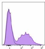
Spark Blue™ 550 anti-human HLA-DR (Flexi-Fluor™)
-
Spark Red™ 718 anti-human HLA-DR (Flexi-Fluor™)


Follow Us