 Login / Register


- Clone
- BC96 (See other available formats)
- Regulatory Status
- RUO
- Workshop
- V T-072
- Other Names
- Low affinity IL-2R, IL-2R α chain, Tac, p55
- Isotype
- Mouse IgG1, κ
- Ave. Rating
- Submit a Review
- Product Citations
- publications

-

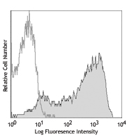
PHA-stimulated (3 day) human peripheral blood lymphocytes were stained with CD25 (clone BC96) PE (solid line) or mouse IgG1, κ PE isotype control (dotted line).
| Cat # | Size | Price | Quantity Check Availability | Save | ||
|---|---|---|---|---|---|---|
| 302605 | 25 tests | 32€ | ||||
| 302606 | 100 tests | 76€ | ||||
CD25 is a 55 kD type I transmembrane glycoprotein also known as the low affinity IL-2 receptor α chain or Tac. It is expressed on progenitor lymphocytes, activated T and B cells, and activated monocytes/macrophages. CD25 is also expressed on a subset of non-stimulated CD4+ T cells termed T regulatory cells. CD25 associates with the IL-2 receptor β (CD122) and common γ chains (CD132) to form the high affinity IL-2R complex.
Product DetailsProduct Details
- Verified Reactivity
- Human
- Reported Reactivity
- Baboon, Chimpanzee, Cynomolgus, Pigtailed Macaque, Rhesus
- Antibody Type
- Monoclonal
- Host Species
- Mouse
- Formulation
- Phosphate-buffered solution, pH 7.2, containing 0.09% sodium azide and BSA (origin USA)
- Preparation
- The antibody was purified by affinity chromatography, and conjugated with PE under optimal conditions.
- Concentration
- Lot-specific (to obtain lot-specific concentration and expiration, please enter the lot number in our Certificate of Analysis online tool.)
- Storage & Handling
- The CD25 antibody solution should be stored undiluted between 2°C and 8°C, and protected from prolonged exposure to light. Do not freeze.
- Application
-
FC - Quality tested
- Recommended Usage
-
Each lot of this antibody is quality control tested by immunofluorescent staining with flow cytometric analysis. For flow cytometric staining, the suggested use of this reagent is 5 µl per million cells in 100 µl staining volume or 5 µl per 100 µl of whole blood.
- Excitation Laser
-
Blue Laser (488 nm)
Green Laser (532 nm)/Yellow-Green Laser (561 nm)
- Application Notes
-
Additional reported applications include: immunocytochemistry3.
- Application References
- Product Citations
-
- RRID
-
AB_314275 (BioLegend Cat. No. 302605)
AB_314276 (BioLegend Cat. No. 302606)
Antigen Details
- Structure
- Type I transmembrane glycoprotein, 55 kD
- Distribution
-
Activated T cells and B cells, monocytes/macrophages, Treg
- Function
- Associates with IL-2 receptor β (CD122) and γ chains (CD132) to form high affinity IL-2R complex
- Ligand/Receptor
- IL-2
- Cell Type
- B cells, Macrophages, Monocytes, T cells, Tregs
- Biology Area
- Cell Biology, Immunology, Neuroscience, Neuroscience Cell Markers
- Molecular Family
- CD Molecules, Cytokine/Chemokine Receptors
- Antigen References
-
1. Taniguchi T, et al. 1993. Cell 73:5.
2. Waldmann T. 1991. J. Biol. Chem. 266:2681. - Gene ID
- 3559 View all products for this Gene ID
- UniProt
- View information about CD25 on UniProt.org
Related Pages & Pathways
Pathways


Related FAQs
- What type of PE do you use in your conjugates?
- We use R-PE in our conjugates.
Other Formats
View All CD25 Reagents Request Custom ConjugationCustomers Also Purchased
Compare Data Across All Formats
This data display is provided for general comparisons between formats.
Your actual data may vary due to variations in samples, target cells, instruments and their settings, staining conditions, and other factors.
If you need assistance with selecting the best format contact our expert technical support team.
-
APC anti-human CD25

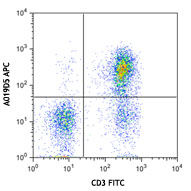
PHA-stimulated (3 day) human peripheral blood lymphocytes we... -
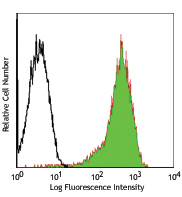
FITC anti-human CD25

PHA-stimulated (3 day) human peripheral blood lymphocytes we... -
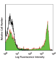
PE anti-human CD25

PHA-stimulated (3 day) human peripheral blood lymphocytes we... -
PE/Cyanine5 anti-human CD25

PHA-stimulated (3 day) human peripheral blood lymphocytes we... -
Purified anti-human CD25

PHA-stimulated (3 day) human peripheral blood lymphocytes we... -
APC/Cyanine7 anti-human CD25

PHA-stimulated (3 day) human peripheral blood lymphocytes we... -
PE/Cyanine7 anti-human CD25

PHA-stimulated (3 day) human peripheral blood lymphocytes we... -
Alexa Fluor® 488 anti-human CD25

PHA-stimulated (3 day) human peripheral blood lymphocytes we... -
Alexa Fluor® 647 anti-human CD25

PHA-stimulated (3 day) human peripheral blood lymphocytes we... -
Pacific Blue™ anti-human CD25

PHA-stimulated (3 day) human peripheral blood lymphocytes we... -
Alexa Fluor® 700 anti-human CD25

PHA-stimulated (3 day) human peripheral blood lymphocytes we... -
Biotin anti-human CD25

PHA-stimulated (3 day) human peripheral blood lymphocytes we... -
PerCP/Cyanine5.5 anti-human CD25

PHA-stimulated (day 3) human peripheral blood lymphocytes we... -
Brilliant Violet 421™ anti-human CD25

Human peripheral blood lymphocytes were stained with CD4 FIT... 
-
Brilliant Violet 605™ anti-human CD25

Human peripheral blood lymphocytes were stained with CD4 FIT... -
Brilliant Violet 650™ anti-human CD25

Human peripheral blood lymphocytes were stained with CD4 FIT... 
-
Brilliant Violet 711™ anti-human CD25

Human peripheral blood lymphocytes were stained with CD4 FIT... -
Brilliant Violet 785™ anti-human CD25

Human peripheral blood lymphocytes were stained with CD4 FIT... -
Brilliant Violet 510™ anti-human CD25

PHA-stimulated (3 day) human peripheral blood lymphocytes we... -
APC/Fire™ 750 anti-human CD25

PHA-stimulated (day 4) human peripheral blood lymphocytes we... 
-
TotalSeq™-A0085 anti-human CD25
-
PE/Dazzle™ 594 anti-human CD25

PHA-stimulated (3 day) human peripheral blood lymphocytes we... -
TotalSeq™-B0085 anti-human CD25
-
TotalSeq™-C0085 anti-human CD25
-
TotalSeq™-D0085 anti-human CD25


Follow Us