 Login / Register


- Clone
- 29A1.4 (See other available formats)
- Regulatory Status
- RUO
- Other Names
- NKp46, NCR1
- Isotype
- Rat IgG2a, κ
- Ave. Rating
- Submit a Review
- Product Citations
- publications

-

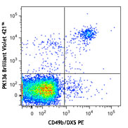
C57BL/6 splenocytes stained with NK1.1 APC and NKp46 (clone 29A1.4) Brilliant Violet 421™ (top) or rat IgG2a Brilliant Violet 421™ isotype control (bottom). -

| Cat # | Size | Price | Quantity Check Availability | Save | ||
|---|---|---|---|---|---|---|
| 137611 | 125 µL | 164€ | ||||
| 137612 | 50 µg | 194€ | ||||
CD335, also known as NKp46, is a single-pass type I membrane protein of 46 kD. It belongs to the natural cytotoxicity receptor (NCR) family and contains two Ig-like (immunoglobulin-like) domains. It's expression is restricted to NK cells and a subset of NKT cells; it's not expressed in CD1d-restricted NKT cells. CD335 is a receptor for viral hemagglutinins and heparan sulfate proteoglycans and is involved in NK cell activation.
Product DetailsProduct Details
- Verified Reactivity
- Mouse
- Antibody Type
- Monoclonal
- Host Species
- Rat
- Immunogen
- NKP46-IgG1 Fc fusion protein
- Formulation
- Phosphate-buffered solution, pH 7.2, containing 0.09% sodium azide and BSA (origin USA).
- Preparation
- The antibody was purified by affinity chromatography and conjugated with Brilliant Violet 421™ under optimal conditions.
- Concentration
- µg sizes: 0.2 mg/mLµL sizes: lot-specific (to obtain lot-specific concentration and expiration, please enter the lot number in our Certificate of Analysis online tool.)
- Storage & Handling
- The antibody solution should be stored undiluted between 2°C and 8°C, and protected from prolonged exposure to light. Do not freeze.
- Application
-
FC - Quality tested
- Recommended Usage
-
Each lot of this antibody is quality control tested by immunofluorescent staining with flow cytometric analysis. For immunofluorescent staining using the µg size, the suggested use of this reagent is ≤0.5 µg per million cells in 100 µl volume. For immunofluorescent staining using the µl size, the suggested use of this reagent is 5 µl per million cells in 100 µl staining volume or 5 µl per 100 µl of whole blood. It is recommended that the reagent be titrated for optimal performance for each application.
Brilliant Violet 421™ excites at 405 nm and emits at 421 nm. The standard bandpass filter 450/50 nm is recommended for detection. Brilliant Violet 421™ is a trademark of Sirigen Group Ltd.
Learn more about Brilliant Violet™.
This product is subject to proprietary rights of Sirigen Inc. and is made and sold under license from Sirigen Inc. The purchase of this product conveys to the buyer a non-transferable right to use the purchased product for research purposes only. This product may not be resold or incorporated in any manner into another product for resale. Any use for therapeutics or diagnostics is strictly prohibited. This product is covered by U.S. Patent(s), pending patent applications and foreign equivalents. - Excitation Laser
-
Violet Laser (405 nm)
- Application Notes
-
Additional reported applications (for the relevant formats) include: immunohistochemical staining of frozen tissue sections1,2 and in vitro activation of NK cells1.
- Application References
-
- Walzer T, et al. 2007. P. Natl. Acad. Sci. USA 104:3384. (FC, Activ)
- Walzer T, et al. 2007. Nat. Immunol. 8:1337. (FC, Activ)
- Guerriero JL, et al. 2011. J. Immunol. 186:3517. (IHC) PubMed
- Product Citations
-
- RRID
-
AB_10915472 (BioLegend Cat. No. 137611)
AB_2563104 (BioLegend Cat. No. 137612)
Antigen Details
- Structure
- Single-pass type I membrane protein, 46 kD; belongs to the natural cytotoxicity receptor (NCR) family; contains 2 Ig-like (immunoglobulin-like) domains
- Distribution
-
Mature and immature NK cells, subset of NKT cells, but not on CD1d-restricted NKT cells
- Function
- NK cells activation
- Ligand/Receptor
- Viral hemagglutinins, heparan sulfate proteoglycans
- Cell Type
- NK cells, NKT cells
- Biology Area
- Immunology, Innate Immunity
- Molecular Family
- CD Molecules
- Antigen References
-
1. Colucci F and Cilio CM. 2010. Nat. Immunol. 125:60.
2. Caligiuri MA. 2008. Blood 112:461.
3. Colonna M. 2009. Immunity 31:15. - Gene ID
- 17086 View all products for this Gene ID
- UniProt
- View information about CD335 on UniProt.org
Related Pages & Pathways
Pathways


Related FAQs
- What is the F/P ratio range of our BV421™ format antibody reagents?
-
It is lot-specific. On average it ranges between 2-4.
Other Formats
View All CD335 Reagents Request Custom ConjugationCustomers Also Purchased
Compare Data Across All Formats
This data display is provided for general comparisons between formats.
Your actual data may vary due to variations in samples, target cells, instruments and their settings, staining conditions, and other factors.
If you need assistance with selecting the best format contact our expert technical support team.
-
Brilliant Violet 510™ anti-mouse CD335 (NKp46)

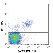
C57BL/6 splenocytes were stained with NK1.1 (clone PK136) AP... 
-
Brilliant Violet 711™ anti-mouse CD335 (NKp46)

C57BL/6 splenocytes were stained with NK1.1 (clone PK136) AP... 
-
PE anti-mouse CD335 (NKp46)

C57BL/6 mouse splenocytes stained with 29A1.4 PE and NK1.1 (... -
PE/Cyanine7 anti-mouse CD335 (NKp46)

C57/BL6 mouse splenocytes were stained with NK1.1 Brilliant ... 
-
Purified anti-mouse CD335 (NKp46)
C57BL/6 mouse splenocytes stained with purified 29A1.4 conju... -
FITC anti-mouse CD335 (NKp46)

C57BL/6 splenocytes stained with NK1.1 (PK136) APC and 29A1.... 
C57BL/6 splenocytes stained with NK1.1 (PK136) APC and rat I... -
APC anti-mouse CD335 (NKp46)

C57BL/6 mouse splenocytes stained with NK1.1 (PK136) PE and ... 
C57BL/6 mouse splenocytes stained with NK1.1 (PK136) PE and ... -
PerCP/Cyanine5.5 anti-mouse CD335 (NKp46)

C57BL/6 splenocytes stained with NK-1.1 (PK136) FITC and Per... 
C57BL/6 splenocytes stained with NK-1.1 (PK136) FITC and Per... -
Brilliant Violet 421™ anti-mouse CD335 (NKp46)

C57BL/6 splenocytes stained with NK1.1 APC and NKp46 (clone ... 
-
Biotin anti-mouse CD335 (NKp46)

C57/BL6 mouse splenocytes were stained with NK1.1 Brilliant ... 
-
Brilliant Violet 605™ anti-mouse CD335 (NKp46)

C57BL/6 splenocytes stained with NK1.1 PE and NKp46 (clone 2... 
-
Purified anti-mouse CD335 (NKp46) (Maxpar® Ready)

Mouse splenocytes stained with 153Eu-anti-CD335 (29A1.4) and... -
Alexa Fluor® 647 anti-mouse CD335 (NKp46)

C57BL/6 mouse splenocytes were stained with NK1.1 FITC and C... 
-
PE/Dazzle™ 594 anti-mouse CD335 (NKp46)

C57BL/6 splenocytes were stained with NK1.1 FITC and CD335 (... 
-
APC/Fire™ 750 anti-mouse CD335 (NKp46)

C57BL/6 splenocytes were stained with NK1.1 PE and CD335 (cl... 
-
Brilliant Violet 650™ anti-mouse CD335 (NKp46)

C57BL/6 splenocytes stained with NK1.1 (clone PK136) FITC an... -
TotalSeq™-A0184 anti-mouse CD335 (NKp46)
-
Brilliant Violet 785™ anti-mouse CD335 (NKp46)

C57BL/6 splenocytes stained with NK1.1 (clone PK136) FITC an... -
Ultra-LEAF™ Purified anti-mouse CD335 (NKp46)
C57BL/6 mouse splenocytes stained with purified 29A1.4 conju... -
TotalSeq™-B0184 anti-mouse CD335 (NKp46)
-
TotalSeq™-C0184 anti-mouse CD335 (NKp46)
-
APC/Cyanine7 anti-mouse CD335 (NKp46)

C57BL/6 mouse splenocytes were stained with NK1.1 Alexa Fluo... -
PE/Cyanine5 anti-mouse CD335 (NKp46)

C57BL/6 splenocytes were stained with anti-mouse NK-1.1 FITC... -
PerCP/Fire™ 806 anti-mouse CD335 (NKp46)

C57BL/6 mouse splenocytes were stained with anti-mouse NK1.... -
PerCP/Fire™ 780 anti-mouse CD335 (NKp46)

C57BL/6 mouse splenocytes were stained with anti-mouse NK1.1... -
Spark Red™ 718 anti-mouse CD335 (NKp46) (Flexi-Fluor™)


Follow Us