 Login / Register


- Clone
- AD2 (See other available formats)
- Regulatory Status
- RUO
- Workshop
- V B-CD73.3
- Other Names
- Ecto-5'-nucleotidase, E.C3.1.3.5, L-VAP-2, NT5E, 5'-NT
- Isotype
- Mouse IgG1, κ
- Ave. Rating
- Submit a Review
- Product Citations
- publications

-

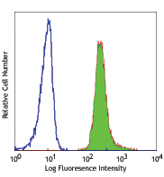
Human peripheral blood lymphocytes were stained with CD3 FITC and CD73 (clone AD2) Brilliant Violet 421™ or mouse IgG1, κ Brilliant Violet 421™ isotype control (bottom). -

Frozen human tonsil section was fixed with 4% paraformaldehyde (PFA) for ten minutes at room temperature and blocked with 5% FBS for 30 minutes at room temperature. Then the section was stained with 10 µg/ml of Brilliant Violet 421™ anti-human CD73 (clone AD2) (blue), 10 µg/ml of Alexa Fluor® 647 anti-human CD19 (clone HIB19) (red) and 10 µg/ml of Alexa Fluor® 488 anti-human CD3 (clone UCHT1) overnight at 4°C. The image was captured by 10X objective. -

| Cat # | Size | Price | Quantity Check Availability | Save | ||
|---|---|---|---|---|---|---|
| 344007 | 25 tests | 152€ | ||||
| 344008 | 100 tests | 305€ | ||||
CD73 is a 70 kD glycophosphatidylinositol (GPI)-linked 5'-nucleotidase, which is also known as ecto-5'-nucleotidase. It converts adenosine monophosphate (AMP) to adenosine. CD73 is expressed on subsets of T and B cells, mesenchymal stem cells, follicular dendritic cells, endothelial cells, and epithelial cells. It has been reported that CD73 costimulates T cell activation, and mediates adhesion of lymphocytes to follicular dendritic cells and endothelial cells.
Product DetailsProduct Details
- Verified Reactivity
- Human
- Reported Reactivity
- African Green, Baboon
- Antibody Type
- Monoclonal
- Host Species
- Mouse
- Formulation
- Phosphate-buffered solution, pH 7.2, containing 0.09% sodium azide and BSA (origin USA).
- Preparation
- The antibody was purified by affinity chromatography and conjugated with Brilliant Violet 421™ under optimal conditions.
- Concentration
- Lot-specific (to obtain lot-specific concentration and expiration, please enter the lot number in our Certificate of Analysis online tool.)
- Storage & Handling
- The antibody solution should be stored undiluted between 2°C and 8°C, and protected from prolonged exposure to light. Do not freeze.
- Application
-
FC - Quality tested
IHC-F - Verified - Recommended Usage
-
Each lot of this antibody is quality control tested by immunofluorescent staining with flow cytometric analysis. For flow cytometric staining, the suggested use of this reagent is 5 µl per million cells in 100 µl staining volume or 5 µl per 100 µl of whole blood. For immunohistochemical staining on frozen tissue sections, the suggested use of this reagent is 5.0 - 10 µg per ml. It is recommended that the reagent be titrated for optimal performance for each application.
Brilliant Violet 421™ excites at 405 nm and emits at 421 nm. The standard bandpass filter 450/50 nm is recommended for detection. Brilliant Violet 421™ is a trademark of Sirigen Group Ltd.
Learn more about Brilliant Violet™.
This product is subject to proprietary rights of Sirigen Inc. and is made and sold under license from Sirigen Inc. The purchase of this product conveys to the buyer a non-transferable right to use the purchased product for research purposes only. This product may not be resold or incorporated in any manner into another product for resale. Any use for therapeutics or diagnostics is strictly prohibited. This product is covered by U.S. Patent(s), pending patent applications and foreign equivalents. - Excitation Laser
-
Violet Laser (405 nm)
- Application Notes
-
Additional reported applications (for the relevant formats) include:immunofluorescence3.
Clone AD2 has been noted to induce clustering and internalization of CD73 in vivo and inhibit metastasis in a murine breast cancer xenograft model4. - Application References
-
- Nakamura T, et al. 1993. J. Immunol. 151:6933.
- Liao J, et al. 2011. J Endod. 37:1217. PubMed
- Touboul C, et al. 2013. J. Transl. Med. 11:28. (IF)
- Terp MG, et al. 2013. J Immunol. 191: 4165-73 (Block)
- Product Citations
-
- RRID
-
AB_10900981 (BioLegend Cat. No. 344007)
AB_11204424 (BioLegend Cat. No. 344008)
Antigen Details
- Structure
- GPI-linked 5'-nucleotidase, 70 kD
- Distribution
-
Subsets of T cells and B cells, mesenchymal stem cells, follicular dendritic cells, endothelial cells, and epithelial cells
- Function
- Catalyses dephosphorylation of adenosine monophosphate, costimulates T cell activation, mediates adhesion of lymphocytes to follicular dendritic cells and endothelial cells
- Cell Type
- B cells, Dendritic cells, Endothelial cells, Epithelial cells, Mesenchymal Stem Cells, T cells, Tregs
- Biology Area
- Costimulatory Molecules, Immunology, Stem Cells
- Molecular Family
- Adhesion Molecules, CD Molecules
- Antigen References
-
1. Zola H, et al. 2007. Leukocyte and stromal Cell Molecules:the CD Markers. A John Wiley & Sons Inc, Publication.
2. Airas L and Jalkanen S, et al. 1996. Blood 88:1755.
3. Gutensohn W, et al. 1995. Cell Immunol. 161:213.
4. Airas L, et al. 1995. J. Exp. Med. 182:1603. - Gene ID
- 4907 View all products for this Gene ID
- UniProt
- View information about CD73 on UniProt.org
Related Pages & Pathways
Pathways


Related FAQs
- What is the F/P ratio range of our BV421™ format antibody reagents?
-
It is lot-specific. On average it ranges between 2-4.
Other Formats
View All CD73 Reagents Request Custom ConjugationCustomers Also Purchased
Compare Data Across All Formats
This data display is provided for general comparisons between formats.
Your actual data may vary due to variations in samples, target cells, instruments and their settings, staining conditions, and other factors.
If you need assistance with selecting the best format contact our expert technical support team.
-
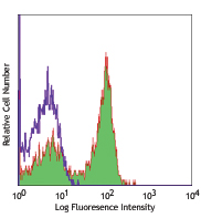
FITC anti-human CD73 (Ecto-5'-nucleotidase)

Human peripheral blood lymphocytes were stained with CD3 APC... 
-
Brilliant Violet 421™ anti-human CD73 (Ecto-5'-nucleotidase)

Human peripheral blood lymphocytes were stained with CD3 FIT... 

Frozen human tonsil section was fixed with 4% paraformaldehy... -
Purified anti-human CD73 (Ecto-5'-nucleotidase)

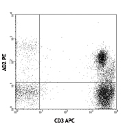
Human peripheral blood lymphocytes stained with AD2 PE and C... 
Human peripheral blood lymphocytes stained with AD2 PE and C... 
Frozen human tonsil section was fixed with 4% paraformaldehy... -
PE anti-human CD73 (Ecto-5'-nucleotidase)
Human peripheral blood lymphocytes stained with AD2 PE and C...
Human peripheral blood lymphocytes stained with AD2 PE and C... -
APC anti-human CD73 (Ecto-5'-nucleotidase)

Human peripheral blood lymphocytes stained with AD2 APC and ... 
-
PE/Cyanine7 anti-human CD73 (Ecto-5'-nucleotidase)

Human peripheral blood lymphocytes were stained with CD3 Pac... -
Pacific Blue™ anti-human CD73 (Ecto-5'-nucleotidase)

Human peripheral blood lymphocytes were stained with CD3 FIT... 
-
PerCP/Cyanine5.5 anti-human CD73 (Ecto-5'-nucleotidase)

Human peripheral blood lymphocytes were stained with CD3 APC... -
Biotin anti-human CD73 (Ecto-5'-nucleotidase)

Human peripheral blood lymphocytes were stained CD3 APC and ... 
-
PE/Dazzle™ 594 anti-human CD73 (Ecto-5'-nucleotidase)

Human peripheral blood lymphocytes were stained with CD3 APC... 
-
APC/Cyanine7 anti-human CD73 (Ecto-5'-nucleotidase)

Human peripheral blood lymphocytes were stained with CD3 FIT... -
Brilliant Violet 605™ anti-human CD73 (Ecto-5'-nucleotidase)

Human peripheral blood lymphocytes were stained with CD3 FIT... 
-
Brilliant Violet 711™ anti-human CD73 (Ecto-5'-nucleotidase)

Human peripheral blood lymphocytes were stained with CD3 PE ... 
-
Brilliant Violet 785™ anti-human CD73 (Ecto-5'-nucleotidase)

Human peripheral blood lymphocytes were stained with CD3 PE ... 
-
TotalSeq™-A0577 anti-human CD73 (Ecto-5'-nucleotidase)
-
TotalSeq™-C0577 anti-human CD73 (Ecto-5'-nucleotidase)
-
TotalSeq™-B0577 anti-human CD73 (Ecto-5'-nucleotidase)
-
APC/Fire™ 750 anti-human CD73 (Ecto-5'-nucleotidase)

Human peripheral blood lymphocytes were stained with anti-hu... -
TotalSeq™-D0577 anti-human CD73 (Ecto-5'-nucleotidase)
-
Alexa Fluor® 700 anti-human CD73 (Ecto-5'-nucleotidase)

Human peripheral blood lymphocytes were stained with anti-hu... -
Alexa Fluor® 647 anti-human CD73 (Ecto-5'-nucleotidase)

Human peripheral blood lymphocytes were stained with anti-hu... -
Brilliant Violet 510™ anti-human CD73 (Ecto-5'-nucleotidase)

Human peripheral blood lymphocytes were stained with anti-hu... -
PerCP/Fire™ 780 anti-human CD73 (Ecto-5'-nucleotidase)

Human peripheral blood lymphocytes were stained with anti-hu... -
Spark Red™ 718 anti-hu CD73 (Ecto-5'-nucleotidase) (Flexi-Fluor™)


Follow Us