 Login / Register


- Clone
- 6H2.B4 (See other available formats)
- Regulatory Status
- RUO
- Other Names
- Cyt c
- Isotype
- Mouse IgG1, κ
- Ave. Rating
- Submit a Review
- Product Citations
- publications

-

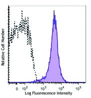
C57BL/6 splenocytes were treated with BioLegend's Fixation Buffer and Permeabilization Wash Buffer, and then were stained with Cytochrome C (clone 6H2-B4) Alexa Fluor® 488 (filled histogram) or mouse IgG1, κ Alexa Fluor® 488 isotype control (open histogram).
| Cat # | Size | Price | Quantity Check Availability | Save | ||
|---|---|---|---|---|---|---|
| 612308 | 100 µg | 212€ | ||||
Cytochrome c is a 15 kD protein found in the mitochondrial intermembrane space with a heme-binding domain. Cytochrome c is a component of the electron transport chain; the heme group transfers electrons from cytochrome b-c1 complex to cytochrome oxidase complex. Cytochrome c initiates apoptosis by release to cytoplasm and binding Apaf-1 which activates procaspase 9. Cytochrome c interacts with the cytochrome b-c1 complex, cytochrome oxidase complex, heme, Apaf-1, and Caspase 9 proteins. The 6H2.B4 monoclonal antibody recognizes human, mouse, and rat cytochrome-c and has been shown to be useful for intracellular flow cytometric staining, Western blotting, immunoprecipitation, and immunofluorescence staining.
Product DetailsProduct Details
- Verified Reactivity
- Mouse, Rat, Human
- Antibody Type
- Monoclonal
- Host Species
- Mouse
- Immunogen
- Rat cyt c-OVA
- Formulation
- Phosphate-buffered solution, pH 7.2, containing 0.09% sodium azide.
- Preparation
- The antibody was purified by affinity chromatography and conjugated with Alexa Fluor® 488 under optimal conditions.
- Concentration
- 0.5 mg/ml
- Storage & Handling
- The antibody solution should be stored undiluted between 2°C and 8°C, and protected from prolonged exposure to light. Do not freeze.
- Application
-
ICFC - Quality tested
- Recommended Usage
-
Each lot of this antibody is quality control tested by intracellular immunofluorescent staining with flow cytometric analysis. For flow cytometric staining, the suggested use of this reagent is ≤0.5 µg per million cells in 100 µl volume. It is recommended that the reagent be titrated for optimal performance for each application.
* Alexa Fluor® 488 has a maximum emission of 519 nm when it is excited at 488 nm.
Alexa Fluor® and Pacific Blue™ are trademarks of Life Technologies Corporation.
View full statement regarding label licenses - Excitation Laser
-
Blue Laser (488 nm)
- Application Notes
-
Additional reported applications (for the relevant formats) include: intracellular flow cytometry5, immunofluorescence microscopy3,5, immunoprecipitation4, and immunocytochemistry5.
- Application References
-
- Goshorn SC, et al. 1991. J. Biol. Chem. 266:2134.
- Jemmerson R, et al. 1991. Eur. J. Immunol. 21:143.
- Chandra D, et al. 2002. J. Biol. Chem. 277:50842. (IF)
- Semenkova L, et al. 2003. Eur. J. Biochem. 270:4388. (IP)
- Shih S-F, et al. 2001. J. Biol. Chem. 276:21870. (ICFC ICC IF)
- She P, et al. 2011. Am J. Physiol Endcorinol Metab.301:E49. PubMed
- McGuire, KA., et al. 2011. J. Virol 85:10806. PubMed
- Product Citations
-
- RRID
-
AB_2565240 (BioLegend Cat. No. 612308)
Antigen Details
- Structure
- Heme binding domain; 15 kD
- Distribution
-
Mitochondrial intermembrane space
- Function
- Component of electron transport chain; heme group transfers electrons from cytochrome b-c1 complex to cytochrome oxidase complex. Initiates apoptosis by release to cytoplasm and binding Apaf-1 which activates procaspase 9
- Interaction
- Cytochrome b-c1 complex, cytochrome oxidase complex, heme, Apaf-1, Casp9
- Biology Area
- Apoptosis/Tumor Suppressors/Cell Death, Cell Biology, Mitochondrial Function, Neuroscience, Neuroscience Cell Markers
- Molecular Family
- Mitochondrial Markers
- Antigen References
-
1. Liu X, et al. 1996. Cell. 86:147.
2. Li P, et al. 1997. Cell. 91:479.
3. Zhang Z, et al. 2003. Gene 312:61.
4. Ferguson H, et al. 2003. J. Biol. Chem. 278:45793. - Gene ID
- 1355 View all products for this Gene ID
- UniProt
- View information about Cytochrome c on UniProt.org
Related Pages & Pathways
Pathways


Related FAQs
Other Formats
View All Cytochrome c Reagents Request Custom Conjugation| Description | Clone | Applications |
|---|---|---|
| Biotin anti-Cytochrome c | 6H2.B4 | ICFC |
| FITC anti-Cytochrome c | 6H2.B4 | ICFC |
| Purified anti-Cytochrome c | 6H2.B4 | IP,ICC,ICFC |
| Alexa Fluor® 488 anti-Cytochrome c | 6H2.B4 | ICFC |
| Alexa Fluor® 647 anti-Cytochrome c | 6H2.B4 | ICFC,ICC |
| FITC anti-Cytochrome c | 6H2.B4 | FC |
| GMP FITC anti-Cytochrome c | 6H2.B4 | FC |
| TotalSeq™-B0043 anti-Cytochrome c | 6H2.B4 | ICPG |
Customers Also Purchased
Compare Data Across All Formats
This data display is provided for general comparisons between formats.
Your actual data may vary due to variations in samples, target cells, instruments and their settings, staining conditions, and other factors.
If you need assistance with selecting the best format contact our expert technical support team.
-
Biotin anti-Cytochrome c
-
FITC anti-Cytochrome c

Balb/c splenocytes intracellularly stained with 6H2.B4 FITC -
Purified anti-Cytochrome c

Immunoprecipitation/Western Blot analysis using purified ant... 
HeLa cells were fixed with 1% paraformaldehyde (PFA) for 10... -
Alexa Fluor® 488 anti-Cytochrome c

C57BL/6 splenocytes were treated with BioLegend's Fixation B... -
Alexa Fluor® 647 anti-Cytochrome c

C57BL/6 splenocytes were treated with BioLegend's Fixation B... 
A-431 cells were fixed with 4% paraformaldehyde for 10 minut... -
FITC anti-Cytochrome c

Typical quality control results from intracellular staining ... -
GMP FITC anti-Cytochrome c

Typical quality control results from intracellular staining ... -
TotalSeq™-B0043 anti-Cytochrome c


Follow Us