 Login / Register


- Clone
- W19086B (See other available formats)
- Regulatory Status
- RUO
- Other Names
- Erythroid 2 Like 2, NF-E2-Related Factor 2, HEBP1, IMDDHH
- Isotype
- Rat IgG2a, κ
- Ave. Rating
- Submit a Review
- Product Citations
- publications

-

Whole cell lysates (15 µg) from untreated (-) or MG-132 treated (+) (10 µM, 10 hours) HeLa cells were resolved by 4-12% Bis-Tris gel electrophoresis, transferred to a PVDF membrane and probed with 1.0 µg/mL (1:500 dilution) of purified anti-NRF2 antibody (clone W19086B) overnight at 4°C. Proteins were visualized by chemiluminescence detection using HRP goat anti-rat IgG antibody (Cat. No. 405422) at a 1:3000 dilution. Direct-Blot™ HRP anti-GAPDH antibody (Cat. No. 607904) was used as a loading control at a 1:50000 dilution (lower). Lane M: Molecular weight marker. -

Untreated (panel A) or MG-132 treated (panel B) (10 µM, 10 hours) HeLa cells were fixed with 4% paraformaldehyde for 10 minutes, permeabilized with methanol for 10 minutes, and blocked with 5% FBS for 30 minutes. Cells were then intracellularly stained with a 1.0 µg/mL (1:500 dilution) of purified anti-NRF2 antibody (clone W19086B) overnight at 4°C, followed by incubation with Alexa Fluor® 594 Goat anti-rat IgG antibody (Cat. No. 405422) at 2.5 µg/mL. Nuclei were counterstained with DAPI, and the image was captured with a 60X objective. -

Whole cell extracts (250 µg total protein) prepared from HeLa cells treated with MG-132 (10 µM, 10 hours) were immunoprecipitated overnight with 2.5 µg of purified rat IgG2a, κ isotype ctrl antibody (Cat. No. 400502) or purified anti-NRF2 antibody (clone W19086B). The resulting IP fractions and whole cell extract input (10%) were resolved by 4-12% Bis-Tris gel electrophoresis, transferred to a PVDF membrane and probed with purified anti-NRF2 Antibody (clone W19086B). Lane M: Molecular weight marker. -

IHC staining of purified anti-NRF2 antibody (clone W19086B) on formalin-fixed paraffin-embedded glioblastoma tissues. Following antigen retrieval using sodium citrate H.I.E.R (Cat. No. 928502), the tissue was incubated with 5 µg/mL of the primary antibody overnight at 4°C. BioLegend’s Ultra Streptavidin HRP Kit (Multi-Species, DAB, Cat. No. 929501) was used for detection followed by hematoxylin counterstaining, according to the protocol provided. The image was captured with a 40X objective. -

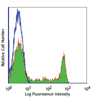
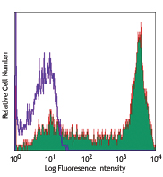
HeLa cells were incubated overnight with (filled histogram) or without (open histogram) the proteasome inhibitor MG132. Cells were fixed with True-Nuclear™ Fix buffer and permeabilized with True-Nuclear™ Perm Buffer. Cells were then stained with purified anti-NRF2 antibody (clone W19086B) followed by anti-rat IgG PE. -

Whole cell lysates (15 μg) from untreated (-) or MG-132 treated (+) (10 μM, 10 hours) NIH/3T3 cells were resolved by 4-12% Bis-Tris gel electrophoresis, transferred to a PVDF membrane and probed with 1.0 μg/mL (1:500 dilution) of purified anti-NRF2 antibody (clone W19086B) overnight at 4°C. Proteins were visualized by chemiluminescence detection using HRP goat anti-rat IgG antibody (Cat. No. 405405) at a 1:3000 dilution. Direct-Blot™ HRP anti-GAPDH antibody (Cat. No. 607904) was used as a loading control at a 1:50000 dilution (lower). Lane M: Molecular weight marker
| Cat # | Size | Price | Quantity Check Availability | Save | ||
|---|---|---|---|---|---|---|
| 939201 | 25 µg | 109€ | ||||
| 939202 | 100 µg | 268€ | ||||
NRF2 is a transcription factor that plays a critical role in inducing expression of genes required for oxidative stress defense and stress balance. It does so by binding to antioxidant response elements (AREs), which are located upstream of target genes. NRF2 is degraded by ubiquitination of KEAP1 E3 ligase. In a clinical setting, NRF2 is frequently activated in many types of cancers; it drives metabolic adaptation and survival in ROS-rich tumor microenvironments.
Product DetailsProduct Details
- Verified Reactivity
- Human, Mouse
- Antibody Type
- Monoclonal
- Host Species
- Rat
- Immunogen
- Partial recombinant human NRF2 protein
- Formulation
- Phosphate-buffered solution, pH 7.2, containing 0.09% sodium azide
- Preparation
- The antibody was purified by affinity chromatography.
- Concentration
- 0.5 mg/mL
- Storage & Handling
- The antibody solution should be stored undiluted between 2°C and 8°C.
- Application
-
WB - Quality tested
ICC, IP, IHC-P, ICFC - Verified - Recommended Usage
-
Each lot of this antibody is quality control tested by western blotting. For western blotting, the suggested use of this reagent is 1.0 µg/mL. For immunocytochemistry, a concentration range of 1.0 - 5.0 μg/mL is recommended. For immunoprecipitation, the suggested use of this reagent is 2.5 µg/test. For immunohistochemistry, a concentration range of 5.0 - 20.0 µg/mL is suggested. For intracellular flow cytometric staining, the suggested use of this reagent is ≤ 0.125 µg per million cells in 100 µL volume. It is recommended that the reagent be titrated for optimal performance for each application.
- Application Notes
-
This clone was tested for ICC on 4% PFA-fixed HeLa cells and permeabilized with either methanol or Triton X-100. Both permeabilization methods were compatible with NRF2 staining.
Clone W19086B is validated to detect NRF2 in mouse NIH/3T3 cells using a Western blot on whole cell lysate. - Product Citations
-
- RRID
-
AB_2892502 (BioLegend Cat. No. 939201)
AB_2892502 (BioLegend Cat. No. 939202)
Antigen Details
- Structure
- NRF2 is a 605 amino acid protein with a predicted molecular weight of 67 kD.
- Distribution
-
Ubiquitously expressed/Nucleus
- Function
- Transcription factor/Redox balance
- Biology Area
- Cell Biology, Mitochondrial Function, Transcription Factors
- Antigen References
-
- M. da Costa R, et al. 2019. Front In Pharm. 10:3389.
- Shoemaker A. 2017. Sci Trans Med. 9:420.
- Robledinos-Antón N, et al. 2019. Hindawi. 10:1155.
- Cuadrado A, et al. 2019. Nat Rev Drug Discov. 18:295-317.
- Gene ID
- 4780 View all products for this Gene ID
- UniProt
- View information about NRF2 on UniProt.org
Related Protocols
- True-Nuclear™ Transcription Factor Staining Protocol for 96-Well U Bottom Plate
- True-Nuclear™ Transcription Factor Staining Protocol for 5mL Tubes
- Immunocytochemistry Staining Protocol
- Western Blotting Protocol
- Immunoprecipitation Protocol
- Immunohistochemistry Protocol for Paraffin-Embedded Sections

Related FAQs
Other Formats
View All NRF2 Reagents Request Custom Conjugation| Description | Clone | Applications |
|---|---|---|
| Purified anti-NRF2 | W19086B | WB,ICC,IP,IHC-P,ICFC |
| PE anti-NRF2 | W19086B | ICFC |
| Alexa Fluor® 647 anti-NRF2 | W19086B | ICFC |
Customers Also Purchased

Compare Data Across All Formats
This data display is provided for general comparisons between formats.
Your actual data may vary due to variations in samples, target cells, instruments and their settings, staining conditions, and other factors.
If you need assistance with selecting the best format contact our expert technical support team.
-
Purified anti-NRF2

Whole cell lysates (15 µg) from untreated (-) or MG-132 trea... 
HeLa cells were incubated overnight with (filled histogram) ... 
Untreated (panel A) or MG-132 treated (panel B) (10 µM, 10 h... 
Whole cell extracts (250 µg total protein) prepared from HeL... 
IHC staining of purified anti-NRF2 antibody (clone W19086B) ... 
Whole cell lysates (15 μg) from untreated (-) or MG-132 trea... -
PE anti-NRF2

HeLa cells were incubated overnight with (filled histogram) ... -
Alexa Fluor® 647 anti-NRF2

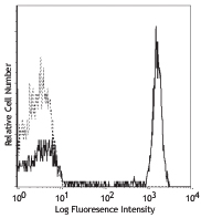
HeLa cells untreated (negative control, open histogram), or ...


Follow Us