 Login / Register


- Clone
- H4A3 (See other available formats)
- Regulatory Status
- RUO
- Workshop
- P PR-63; BP 473; P P008
- Other Names
- Lysosome-Associated Membrane Protein 1, LGP-120, LAMP-1
- Isotype
- Mouse IgG1, κ
- Ave. Rating
- Submit a Review
- Product Citations
- publications

-

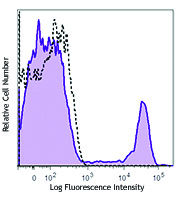
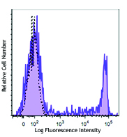
Thrombin-activated human peripheral blood platelets were stained with CD107a (clone H4A3) APC (pink histogram) or mouse IgG1, κ APC (blue histogram).
| Cat # | Size | Price | Quantity Check Availability | Save | ||
|---|---|---|---|---|---|---|
| 328619 | 25 tests | 120€ | ||||
| 328620 | 100 tests | 252€ | ||||
CD107a, also known as Lysosome-Associated Membrane Protein 1 (LAMP-1) or LGP-120, is a 110-140 kD type I membrane glycoprotein. Mature CD107a is heavily glycosylated from a 40 kD core protein. This molecule is located on the luminal side of lysosomes. Upon activation, CD107a is transferred to the cell membrane surface of activated platelets, activated lymphocytes, macrophages, epithelial cells, endothelial cells, and some tumor cells. CD107a has been suggested to play a role in the protection of lysosomal membrane from lysosomal hydrolases which is involved in cell adhesion and regulation of tumor metastasis, and mediates autoimmune disease progression. CD107a is a ligand for galaptin and E-selectin. Surface expression of LAMP-1 has been shown to correlate with CD8+ T cell and NK cell cytotoxicity.
Product DetailsProduct Details
- Verified Reactivity
- Human
- Reported Reactivity
- African Green, Baboon, Chimpanzee, Cynomolgus, Pigtailed Macaque, Rhesus
- Antibody Type
- Monoclonal
- Host Species
- Mouse
- Immunogen
- Human adult adherent peripheral blood cells
- Formulation
- Phosphate-buffered solution, pH 7.2, containing 0.09% sodium azide and BSA (origin USA)
- Preparation
- The antibody was purified by affinity chromatography, and conjugated with APC under optimal conditions.
- Concentration
- Lot-specific (to obtain lot-specific concentration and expiration, please enter the lot number in our Certificate of Analysis online tool.)
- Storage & Handling
- The antibody solution should be stored undiluted between 2°C and 8°C, and protected from prolonged exposure to light. Do not freeze.
- Application
-
FC - Quality tested
- Recommended Usage
-
Each lot of this antibody is quality control tested by immunofluorescent staining with flow cytometric analysis. For flow cytometric staining, the suggested use of this reagent is 5 µl per million cells in 100 µl staining volume or 5 µl per 100 µl of whole blood.
- Excitation Laser
-
Red Laser (633 nm)
- Application Notes
-
Additional reported applications (for the relevant formats) include: Western blotting8, immunohistochemical staining2, immunofluorescence5,7, and immunoprecipitation5.
This antibody is specific to human LAMP-1. Positive control: Hela cells; LAMP-1 molecular weight appears to be at ~110 kDa on the gel due to high glycosylation. - Application References
-
- Misse D, et al. 1999. Blood 93:2454.
- Furuta K, et al. 2001. Am. J. Pathol. 159:449. (IHC)
- Watanabe A, et al. 2011. J. Biol. Chem. 286:10702. PubMed
- Baron Gaillard CL, et al. 2011. Mol. Cell. Biol. 22:5459. PubMed
- Hauck CR and Meyer TF. 1997. FEBS Lett. 405:86. (IF, IP)
- De Keersmaecker B, et al. 2012. J. Virol. 86:9351. PubMed
- Knodler LA, et al. 2010. P. Natl. Acad. Sci. USA. 107:17733. (IF)
- Oh J, et al. 2000. Hum. Mol. Genet. 9:375. (WB)
- Salio M, et al. 2013 PNAS. 110:4753. PubMed
- Product Citations
-
- RRID
-
AB_1279057 (BioLegend Cat. No. 328619)
AB_1279055 (BioLegend Cat. No. 328620)
Antigen Details
- Structure
- LAMP-1 is a 417 amino acid protein with a molecular mass of 45 kD.
- Distribution
-
Macrophages, epithelial cells, endothelial cells, some tumor cells; located on the luminal side of lysosomes or on the surface of cell membranes
- Function
- Protect lysosomal membrane from lysosomal hydrolases, adhesion
- Ligand/Receptor
- Galaptin
- Cell Type
- Endothelial cells, Epithelial cells, Macrophages
- Biology Area
- Cell Biology, Immunology, Neurodegeneration, Neuroscience, Protein Trafficking and Clearance
- Molecular Family
- Adhesion Molecules, CD Molecules
- Antigen References
-
1. Sarafian V, et al. 2006. Arch. Dermatol. Res. 298:7381.
2. Schlossman SF, et al. 1995. Leukocyte Typing V:White Cell Differentiation Antigens. New York:Oxford University Press.
3. Sawada R, et al. 1993. J. Biol. Chem. 268:12675.
4. Chen JW, et al. 1988. J. Biol. Chem. 263:8754.
5. Chen JW, et al. 1986. Biochem. Soc. Symp. 51:97112. - Gene ID
- 3916 View all products for this Gene ID
- UniProt
- View information about CD107a on UniProt.org
Related Pages & Pathways
Pathways


Related FAQs
Other Formats
View All CD107a Reagents Request Custom ConjugationCustomers Also Purchased
Compare Data Across All Formats
This data display is provided for general comparisons between formats.
Your actual data may vary due to variations in samples, target cells, instruments and their settings, staining conditions, and other factors.
If you need assistance with selecting the best format contact our expert technical support team.
-
Biotin anti-human CD107a (LAMP-1)

Thrombin-activated human peripheral blood platelets were sta... -
Purified anti-human CD107a (LAMP-1)

Thrombin-activated human peripheral blood platelets were sta... 
ICC staining of purified anti-LAMP-1 antibody (clone H4A3) o... 
Western blot of purified anti-LAMP-1 antibody (clone H4A3). ... 
Formalin-fixed paraffin-embedded human tonsil treated with a... 
SeqIF™ (sequential immunofluorescence) staining on COMET™ of... -
FITC anti-human CD107a (LAMP-1)

Thrombin-activated human peripheral blood platelets were sta... -
PE anti-human CD107a (LAMP-1)

Thrombin-activated human peripheral blood platelets were sta... -
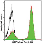
Alexa Fluor® 488 anti-human CD107a (LAMP-1)

Thrombin-activated human peripheral blood platelets were sta... -
Alexa Fluor® 647 anti-human CD107a (LAMP-1)

Thrombin-activated human peripheral blood platelets were sta... -
PerCP/Cyanine5.5 anti-human CD107a (LAMP-1)

PMA+Ionomycin stimulated (4 hours) human peripheral blood mo... -
APC anti-human CD107a (LAMP-1)

Thrombin-activated human peripheral blood platelets were sta... -
Pacific Blue™ anti-human CD107a (LAMP-1)

Thrombin-activated human peripheral blood platelets were sta... -
Brilliant Violet 421™ anti-human CD107a (LAMP-1)

Thrombin-activated human peripheral blood platelets were sta... -
PE/Cyanine7 anti-human CD107a (LAMP-1)

Thrombin-activated human peripheral blood platelets were sta... -
APC/Cyanine7 anti-human CD107a (LAMP-1)

PMA+Ionomycin stimulated (6 hours) human peripheral blood mo... -
Brilliant Violet 510™ anti-human CD107a (LAMP-1)

Thrombin-activated human peripheral blood platelets were sta... -
Brilliant Violet 605™ anti-human CD107a (LAMP-1)

Thrombin-activated human peripheral blood platelets were sta... -
Purified anti-human CD107a (LAMP-1) (Maxpar® Ready)

Human PBMCs were incubated for 5 hours in media alone (top) ... 
-
Brilliant Violet 650™ anti-human CD107a (LAMP-1)

Thrombin-activated human peripheral blood platelets were sta... -
Brilliant Violet 711™ anti-human CD107a (LAMP-1)

Thrombin-activated human peripheral blood platelets were sta... -
PerCP anti-human CD107a (LAMP-1)

Thrombin-activated human peripheral blood platelets were sta... -
Brilliant Violet 785™ anti-human CD107a (LAMP-1)

Thrombin-activated human peripheral blood platelets were sta... -
PE/Dazzle™ 594 anti-human CD107a (LAMP-1)

Thrombin-activated human peripheral blood platelets were sta... -
TotalSeq™-A0155 anti-human CD107a (LAMP-1)
-
TotalSeq™-C0155 anti-human CD107a (LAMP-1)
-
TotalSeq™-B0155 anti-human CD107a (LAMP-1)
-
APC/Fire™ 750 anti-human CD107a (LAMP-1) Antibody

Thrombin-activated human peripheral blood platelets were sta... -
PE/Cyanine5 anti-human CD107a (LAMP-1)

Thrombin-activated human peripheral blood platelets were sta...


Follow Us