 Login / Register


- Clone
- N418 (See other available formats)
- Regulatory Status
- RUO
- Other Names
- αX integrin, integrin αX chain, CR4, p150, ITGAX
- Isotype
- Armenian Hamster IgG
- Ave. Rating
- Submit a Review
- Product Citations
- publications

-

C57BL/6 mouse splenocytes stained with N418 Alexa Fluor® 647 -

Paraformaldehyde-fixed (4%), 500 μm-thick mouse spleen section was processed according to the Ce3DTM Tissue Clearing Kit protocol (cat. no. 427701). The section was costained with anti-mouse/human CD45R/B220 Antibody (clone RA3-6B2) Alexa Fluor® 488 at 5 µg/mL (green), anti-mouse CD8a Antibody (clone 53-6.7) Alexa Fluor® 594 at 5 µg/mL (yellow), and anti-mouse CD11c Antibody (clone N418) Alexa Fluor® 647 at 5 µg/mL (magenta). The section was then optically cleared and mounted in a sample chamber. The image was captured with a 10X objective using Zeiss 780 confocal microscope and processed by Imaris image analysis software.
Watch the video. -

Fixed whole mount mouse epidermis Langerhans cells were stained with Alexa Fluor® 647 CD11c (red) (clone N418) and PE CD207 (yellow) (clone 4C7). Isotype controls at the same concentrations were used for the negative control. Cells were mounted in Prolong Gold and imaged with a Leica SP8 confocal. Image courtesy of Grzegorz Chodaczek and Zbigniew Mikulski at LIAI. -

Live intravital mouse spleen imaging. PE CD11b (red) (clone M1/70), Alexa Fluor® 488 F4/80 (green) (clone BM8), and Alexa Fluor® 647 CD11c (blue) (clone N418) were imaged 30 minutes after iv injection of 10 µg per antibody. Isotype controls at the same concentrations, time post injection, and exposure conditions were used for the negative control. Cells were imaged with a Leica SP5 confocal on anesthetized mice. Image courtesy of Grzegorz Chodaczek and Zbigniew Mikulski at LIAI. -

Fixed whole mount mouse spleen was stained with FITC CD172a (red) (clone P84), Alexa Fluor® CD11c (yellow) (clone N418), and PE CD11b (blue) (clone M1/70). Isotype controls at the same concentrations were used for the negative control. Cells were mounted in Prolong Gold and imaged with a Leica SP8 confocal. Image courtesy of Grzegorz Chodaczek and Zbigniew Mikulski at LIAI. -

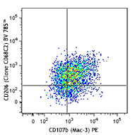
Confocal image of C57BL/6 mouse spleen sample acquired using the IBEX method of highly multiplexed antibody-based imaging: CD11c (cyan) in Cycle 2 and CD68 (red) in Cycle 3. Tissues were prepared using ~1% (vol/vol) formaldehyde and a detergent. Following fixation, samples are immersed in 30% (wt/vol) sucrose for cryoprotection. Images are courtesy of Drs. Andrea J. Radtke and Ronald N. Germain of the Center for Advanced Tissue Imaging (CAT-I) in the National Institute of Allergy and Infectious Diseases (NIAID, NIH).
| Cat # | Size | Price | Quantity Check Availability | Save | ||
|---|---|---|---|---|---|---|
| 117314 | 25 µg | 81€ | ||||
| 117312 | 100 µg | 184€ | ||||
CD11c is a 150 kD glycoprotein also known as αX integrin, CR4, and p150. CD11c forms a αXβ2 heterodimer with β2 integrin (CD18). It is primarily expressed on dendritic cells, NK cells, a subset of intestinal intraepithelial lymphocytes (IEL), and some activated T cells. The αXβ2 integrin plays an important role in cell-cell contact by binding its ligands: iC3b, fibrinogen, and CD54.
Product DetailsProduct Details
- Verified Reactivity
- Mouse
- Antibody Type
- Monoclonal
- Host Species
- Armenian Hamster
- Immunogen
- Mouse spleen dendritic cells
- Formulation
- Phosphate-buffered solution, pH 7.2, containing 0.09% sodium azide
- Preparation
- The antibody was purified by affinity chromatography and conjugated with Alexa Fluor® 647 under optimal conditions.
- Concentration
- 0.5 mg/mL
- Storage & Handling
- The antibody solution should be stored undiluted between 2°C and 8°C, and protected from prolonged exposure to light. Do not freeze.
- Application
-
FC - Quality tested
3D IHC - Verified
IHC - Reported but collaborator, not verified in house
ICC, SB - Reported in the literature, not verified in house - Recommended Usage
-
Each lot of this antibody is quality control tested by immunofluorescent staining with flow cytometric analysis. For flow cytometric staining, the suggested use of this reagent is ≤ 0.25 µg per 106 cells in 100 µL volume. For 3D immunohistochemistry on formalin-fixed tissues, a concentration of 5.0 µg/mL is suggested. It is recommended that the reagent be titrated for optimal performance for other applications.
* Alexa Fluor® 647 has a maximum emission of 668 nm when it is excited at 633nm / 635nm.
Alexa Fluor® and Pacific Blue™ are trademarks of Life Technologies Corporation.
View full statement regarding label licenses - Excitation Laser
-
Red Laser (633 nm)
- Application Notes
-
Additional reported applications (for the relevant formats) include: immunoprecipitation3, immunohistochemical staining of acetone-fixed frozen sections3, immunofluorescence microscopy5, 9 (Alexa Fluor® 488 conjugated N418 was used for IHC in frozen sections10), and spatial biology (IBEX)22,23.
- Additional Product Notes
-
Iterative Bleaching Extended multi-pleXity (IBEX) is a fluorescent imaging technique capable of highly-multiplexed spatial analysis. The method relies on cyclical bleaching of panels of fluorescent antibodies in order to image and analyze many markers over multiple cycles of staining, imaging, and, bleaching. It is a community-developed open-access method developed by the Center for Advanced Tissue Imaging (CAT-I) in the National Institute of Allergy and Infectious Diseases (NIAID, NIH).
- Application References
-
- Granucci F, et al. 1997. J. Immunol. 159:1794.
- Stokes RW, et al. 1998. J. Immunol. 160:5514.
- Metlay JP, et al. 1990. J. Exp. Med. 171:1753. (IHC, IP)
- Ma XT, et al. 2006. Cancer Research 66:1169.
- Chin RK, et al. 2006. J. Immunol. 177:290. (IF)
- Cervantes-Barragan L, et al. 2007. Blood 109:1131. (FC) PubMed
- Turnquist HR, et al. 2007. J. Immunol. 178:7018. (FC) PubMed
- Benson MJ, et al. 2007. J. Exp. Med. doi:10.1084/jem.20070719. (FC) PubMed
- You Y, et al. 2009. J. Immunol. 182:7343. (IF) PubMed
- Roland CL, et al. 2009. Mol. Cancer Res. 8:1761. (IHC, FC) PubMed
- Wikstrom M, et al.2006. J. Immunol. 177:913. PubMed
- Pericolini E, et al. 2008. J. Leukocyte Biol. 83:1286. PubMed
- Randall LM, et al. 2008. Infect. Immun.76:3312. PubMed
- Fahlen-Yrild L, et al. 2009. J. Immunol. 183:5032. PubMed
- Osterholzer JJ, et al. 2009. J. Immunol. 183:8044. PubMed
- Bankoti J, et al. 2010. Toxicol. Sci. 115:422. (FC) PubMed
- Eisenach PA, et al. 2010. J Cell Sci. 123:4182. PubMed
- Leppin K, et al. 2014. Invest. Ophthalmol. Vis. Sci. 55:3603. PubMed
- Sakai F, et al. 2014. PLoS One. 9:105370. PubMed
- Gibbins JD, et al. 2014. Blood. 124:2953. PubMed
- White CE, et al. 2015. J Immunol. 194:697. PubMed
- Lu X, et al. 2015. J Immunol. 194:2011. PubMed
- Radtke AJ, et al. 2020. Proc Natl Acad Sci U S A. 117:33455-65. (SB) PubMed
- Radtke AJ, et al. 2022. Nat Protoc. 17:378-401. (SB) PubMed
- Product Citations
-
- RRID
-
AB_492850 (BioLegend Cat. No. 117314)
AB_389328 (BioLegend Cat. No. 117312)
Antigen Details
- Structure
- Integrin α-chain, associates with integrin β2 (CD18), 150 kD
- Distribution
-
Dendritic cells, NK cells, intestinal intraepithelial lymphocytes (IEL), some activated T cells
- Function
- Cellular adhesion
- Ligand/Receptor
- iC3b, fibrinogen
- Cell Type
- Dendritic cells, Epithelial cells, NK cells, T cells, Tregs
- Biology Area
- Cell Adhesion, Cell Biology, Costimulatory Molecules, Immunology, Innate Immunity, Neuroscience, Neuroscience Cell Markers
- Molecular Family
- Adhesion Molecules, CD Molecules
- Antigen References
-
1. Barclay A, et al. 1997. The Leukocyte Antigen Facts Book Academic Press.
2. Springer TA. 1994. Cell 76:301.
3. Lopez-Rodriguez C, et al. 1996. J. Immunol. 156:3780. - Gene ID
- 16411 View all products for this Gene ID
- UniProt
- View information about CD11c on UniProt.org
Related Pages & Pathways
Pathways


Related FAQs
Other Formats
View All CD11c Reagents Request Custom ConjugationCustomers Also Purchased
Compare Data Across All Formats
This data display is provided for general comparisons between formats.
Your actual data may vary due to variations in samples, target cells, instruments and their settings, staining conditions, and other factors.
If you need assistance with selecting the best format contact our expert technical support team.
-
APC anti-mouse CD11c

C57BL/6 mouse splenocytes stained with N418 APC and PK136 PE -
Biotin anti-mouse CD11c

C57BL/6 mouse splenocytes stained with APC anti-mouse I-A/I-... 

Frozen mouse spleen sections. Endogenous biotin blocking app... -
FITC anti-mouse CD11c

C57BL/6 mouse splenocytes stained with APC anti-mouse I-A/I-... 
C57BL/6 mouse splenocytes stained with APC anti-mouse I-A/I-... -
PE anti-mouse CD11c

C57BL/6 mouse splenocytes stained with APC anti-mouse I-A/I-... 
C57BL/6 mouse splenocytes stained with APC anti-mouse I-A/I-... -
Purified anti-mouse CD11c

C57BL/6 mouse splenocytes stained with APC anti-mouse I-A/I-... 
-
Alexa Fluor® 488 anti-mouse CD11c

C57BL/6 mouse splenocytes stained with APC anti-mouse I-A/I-... 
-
Alexa Fluor® 647 anti-mouse CD11c

C57BL/6 mouse splenocytes stained with N418 Alexa Fluor® 647 
Paraformaldehyde-fixed (4%), 500 μm-thick mouse spleen secti... 
Fixed whole mount mouse epidermis Langerhans cells were stai... 
Live intravital mouse spleen imaging. PE CD11b (red) (clone ... 
Confocal image of C57BL/6 mouse spleen sample acquired using... 
Fixed whole mount mouse spleen was stained with FITC CD172a ... -
PE/Cyanine5 anti-mouse CD11c

C57BL/6 mouse splenocytes stained with APC anti-mouse I-A/I-... 
-
PE/Cyanine7 anti-mouse CD11c

C57BL/6 mouse splenocytes stained with APC anti-mouse I-A/I-... 
-
Brilliant Violet 605™ anti-mouse CD11c

C57BL/6 mouse splenocytes were stained with I-A/I-E FITC and... -
Alexa Fluor® 700 anti-mouse CD11c

C57BL/6 mouse splenocytes stained with N418 Alexa Fluor®... -
Pacific Blue™ anti-mouse CD11c

C57BL/6 mouse splenocytes stained with Pacific Blue™ N... -
APC/Cyanine7 anti-mouse CD11c

C57BL/6 mouse splenocytes were stained with I-A/I-E PE and C... -
PerCP/Cyanine5.5 anti-mouse CD11c

C57BL/6 mouse splenocytes stained with N418 PerCP/Cyanine5.5 -
PerCP anti-mouse CD11c

C57BL/6 mouse splenocytes stained with N418 PerCP and M5/114... -
Brilliant Violet 421™ anti-mouse CD11c

C57BL/6 mouse lymph nodes, fixed O/N in PLP, blocked with 10... 

C57BL/6 mouse splenocytes were stained with mouse I-A/I-E FI... -
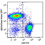
Brilliant Violet 570™ anti-mouse CD11c

C57BL/6 mouse splenocytes were stained with mouse I-A/I-E FI... 
-
Brilliant Violet 785™ anti-mouse CD11c

C57BL/6 mouse splenocytes were stained with I-A/I-E FITC and... -
Brilliant Violet 510™ anti-mouse CD11c

C57BL/6 mouse splenocytes were stained with mouse I-A/I-E AP... 
-
Brilliant Violet 650™ anti-mouse CD11c

C57BL/6 mouse splenocytes were stained with mouse I-A/I-E PE... 
-
Purified anti-mouse CD11c (Maxpar® Ready)

Mouse splenocytes stained with 147Sm-anti-CD45 (30-F11) and ... -
Alexa Fluor® 594 anti-mouse CD11c

C57BL/6 mouse frozen spleen section was fixed with 4% parafo... 
Paraformaldehyde-fixed (4%), 500 μm-thick mouse spleen secti... -
PE/Dazzle™ 594 anti-mouse CD11c

C57BL/6 mouse splenocytes were stained with I-A/I-E FITC and... -
Brilliant Violet 711™ anti-mouse CD11c

C57BL/6 mouse splenocytes were stained with mouse I-A/I-E Pe... -
APC/Fire™ 750 anti-mouse CD11c

C57BL/6 splenocytes were stained with I-A/I-E PE and CD11c (... 
-
TotalSeq™-A0106 anti-mouse CD11c
-
Brilliant Violet 750™ anti-mouse CD11c

C57BL/6 mouse splenocytes were stained with mouse I-A/I-E PE... -
TotalSeq™-B0106 anti-mouse CD11c
-
TotalSeq™-C0106 anti-mouse CD11c
-
KIRAVIA Blue 520™ anti-mouse CD11c

C57BL/6 mouse splenocytes were stained with I-A/I-E APC and ... -
Spark Blue™ 550 anti-mouse CD11c

C57BL/6 mouse splenocytes stained with I-A/I-E (clone M5/114... -
Spark NIR™ 685 anti-mouse CD11c

C57BL/6 mouse splenocytes were stained with anti-mouse I-A/I... -
Spark UV™ 387 anti-mouse CD11c

C57BL/6 mouse splenocytes were stained with anti-mouse I-A/I... -
Spark Red™ 718 anti-mouse CD11c

C57BL/6 mouse splenocytes were stained with anti-mouse I-A/I... -
Spark Blue™ 515 anti-mouse CD11c

C57BL/6 mouse splenocytes were surface stained with anti-mou... -
PerCP/Fire™ 806 anti-mouse CD11c

C57BL/6 splenocytes were stained with anti-mouse I-A/I-E APC...


Follow Us