 Login / Register


- Clone
- SK1 (See other available formats)
- Regulatory Status
- RUO
- Other Names
- T8, Leu2
- Isotype
- Mouse IgG1, κ
- Ave. Rating
- Submit a Review
- Product Citations
- publications

-

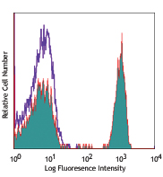
Human peripheral blood lymphocytes stained with SK1 Alexa Fluor® 488 -

Confocal image of human lymph node sample acquired using the IBEX method of highly multiplexed antibody-based imaging: CD1c (red) in Cycle 3, CD8 (blue) in Cycle 4, and CD25 (yellow) in Cycle 4. Tissues were prepared using ~1% (vol/vol) formaldehyde and a detergent. Following fixation, samples are immersed in 30% (wt/vol) sucrose for cryoprotection. Images are courtesy of Drs. Andrea J. Radtke and Ronald N. Germain of the Center for Advanced Tissue Imaging (CAT-I) in the National Institute of Allergy and Infectious Diseases (NIAID, NIH). -

Confocal image of human metastatic lymph node sample acquired using the IBEX method of highly multiplexed antibody-based imaging: CD8 (green) in Cycle 1 and CD138 (blue) in Cycle 5. Tissues were prepared using ~1% (vol/vol) formaldehyde and a detergent. Following fixation, samples are immersed in 30% (wt/vol) sucrose for cryoprotection. Images are courtesy of Drs. Andrea J. Radtke and Ronald N. Germain of the Center for Advanced Tissue Imaging (CAT-I) in the National Institute of Allergy and Infectious Diseases (NIAID, NIH).
| Cat # | Size | Price | Quantity Check Availability | Save | ||
|---|---|---|---|---|---|---|
| 344716 | 100 tests | 259€ | ||||
CD8a is a 32-34 kD type I glycoprotein. It forms a homodimer (CD8a/a) or heterodimer (CD8a/b) with CD8b. CD8, also known as T8 and Leu2, is a member of the immunoglobulin superfamily found on the majority of thymocytes, a subset of peripheral blood T cells, and NK cells (which express almost exclusively CD8a homodimers). CD8 acts as a co-receptor with MHC class I-restricted T cell receptors in antigen recognition and T cell activation and has been shown to play a role in thymic differentiation. Two domains in CD8a are important for function: the extracellular IgSF domain binds the α3 domain of MHC class I and the cytoplasmic CXCP motif binds the tyrosine kinase p56 Lck.
Product DetailsProduct Details
- Verified Reactivity
- Human, Cynomolgus, Rhesus
- Reported Reactivity
- African Green, Chimpanzee, Pigtailed Macaque, Sooty Mangabey
- Antibody Type
- Monoclonal
- Host Species
- Mouse
- Formulation
- Phosphate-buffered solution, pH 7.2, containing 0.09% sodium azide and BSA (origin USA)
- Preparation
- The antibody was purified by affinity chromatography and conjugated with Alexa Fluor® 488 under optimal conditions.
- Concentration
- Lot-specific (to obtain lot-specific concentration, please enter the lot number in our Concentration and Expiration Lookup or Certificate of Analysis online tools.)
- Storage & Handling
- The antibody solution should be stored undiluted between 2°C and 8°C, and protected from prolonged exposure to light. Do not freeze.
- Application
-
FC - Quality tested
SB - Reported in the literature, not verified in house - Recommended Usage
-
Each lot of this antibody is quality control tested by immunofluorescent staining with flow cytometric analysis. For flow cytometric staining, the suggested use of this reagent is 5 µl per million cells in 100 µl staining volume or 5 µl per 100 µl of whole blood.
*Alexa Fluor® 488 has a maximum emission of 519 nm when it is excited at 488 nm.
Alexa Fluor® and Pacific Blue™ are trademarks of Life Technologies Corporation.
View full statement regarding label licenses - Excitation Laser
-
Blue Laser (488 nm)
- Application Notes
-
Clone SK1 recognizes the a chain of CD8. Additional reported applications (for the relevant formats) include: proteogenomics8, immunohistochemistry of acetone-fixed frozen tissue sections, and spatial biology (IBEX)9,10. This clone was tested in-house and does not demonstrate utility for formalin-fixed paraffin-embedded (FFPE) human tonsil sections.
- Additional Product Notes
-
Iterative Bleaching Extended multi-pleXity (IBEX) is a fluorescent imaging technique capable of highly-multiplexed spatial analysis. The method relies on cyclical bleaching of panels of fluorescent antibodies in order to image and analyze many markers over multiple cycles of staining, imaging, and, bleaching. It is a community-developed open-access method developed by the Center for Advanced Tissue Imaging (CAT-I) in the National Institute of Allergy and Infectious Diseases (NIAID, NIH).
- Application References
-
- Ledbetter JA, et al. 1981. J. Exp. Med. 153:310.
- Campanelli R, et al. 2002. Intl. Immunol. 14:39.
- Evans RL, et al. 1981. Immunol. 78:544.
- Wooldridge L, et al. 2005. J. Bio. Chem. 280:27491.
- Ch'el IL, et al. 2011. J Exp Med. 208:633. PubMed
- Carbone A, et al. 1999. Blood 93:2319. (IHC-F)
- Ahmed A, et al. 2001. J. Pathol. 193:383. (IHC)
- Peterson VM, et al. 2017. Nat. Biotechnol. 35:936. (PG)
- Radtke AJ, et al. 2020. Proc Natl Acad Sci USA. 117:33455-33465. (SB) PubMed
- Radtke AJ, et al. 2022. Nat Protoc. 17:378-401. (SB) PubMed
- Product Citations
-
- RRID
-
AB_10549301 (BioLegend Cat. No. 344716)
Antigen Details
- Structure
- Ig superfamily, homodimer or heterodimer with CD8b, 32-34 kD
- Distribution
-
Majority of thymocytes, T cell subset, NK cells
- Function
- MHC class I co-receptor, thymic differentiation, T cell activation
- Ligand/Receptor
- MHC Class I molecules
- Cell Type
- NK cells, T cells, Thymocytes
- Biology Area
- Immunology
- Molecular Family
- CD Molecules
- Antigen References
-
1. Barclay N, et al. 1993. The Leucocyte Antigen FactsBook. Academic Press Inc. San Diego.
- Gene ID
- 925 View all products for this Gene ID
- UniProt
- View information about CD8 on UniProt.org
Related Pages & Pathways
Pathways


Related FAQs
- If an antibody clone has been previously successfully used in IBEX in one fluorescent format, will other antibody formats work as well?
-
It’s likely that other fluorophore conjugates to the same antibody clone will also be compatible with IBEX using the same sample fixation procedure. Ultimately a directly conjugated antibody’s utility in fluorescent imaging and IBEX may be specific to the sample and microscope being used in the experiment. Some antibody clone conjugates may perform better than others due to performance differences in non-specific binding, fluorophore brightness, and other biochemical properties unique to that conjugate.
- Will antibodies my lab is already using for fluorescent or chromogenic IHC work in IBEX?
-
Fundamentally, IBEX as a technique that works much in the same way as single antibody panels or single marker IF/IHC. If you’re already successfully using an antibody clone on a sample of interest, it is likely that clone will have utility in IBEX. It is expected some optimization and testing of different antibody fluorophore conjugates will be required to find a suitable format; however, legacy microscopy techniques like chromogenic IHC on fixed or frozen tissue is an excellent place to start looking for useful antibodies.
- Are other fluorophores compatible with IBEX?
-
Over 18 fluorescent formats have been screened for use in IBEX, however, it is likely that other fluorophores are able to be rapidly bleached in IBEX. If a fluorophore format is already suitable for your imaging platform it can be tested for compatibility in IBEX.
- The same antibody works in one tissue type but not another. What is happening?
-
Differences in tissue properties may impact both the ability of an antibody to bind its target specifically and impact the ability of a specific fluorophore conjugate to overcome the background fluorescent signal in a given tissue. Secondary stains, as well as testing multiple fluorescent conjugates of the same clone, may help to troubleshoot challenging targets or tissues. Using a reference control tissue may also give confidence in the specificity of your staining.
- How can I be sure the staining I’m seeing in my tissue is real?
-
In general, best practices for validating an antibody in traditional chromogenic or fluorescent IHC are applicable to IBEX. Please reference the Nature Methods review on antibody based multiplexed imaging for resources on validating antibodies for IBEX.
Other Formats
View All CD8 Reagents Request Custom ConjugationCustomers Also Purchased
Compare Data Across All Formats
This data display is provided for general comparisons between formats.
Your actual data may vary due to variations in samples, target cells, instruments and their settings, staining conditions, and other factors.
If you need assistance with selecting the best format contact our expert technical support team.
-
Alexa Fluor® 647 anti-human CD8

Human peripheral blood lymphocytes were stained with CD8 (cl... -
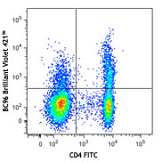
Brilliant Violet 650™ anti-human CD8

Human peripheral blood lymphocytes were stained with CD8 (cl... -
Purified anti-human CD8

Human peripheral blood lymphocytes stained with SK1, followe... -
FITC anti-human CD8
Human peripheral blood lymphocytes stained with SK1 FITC -
PE anti-human CD8

Human peripheral blood lymphocytes stained with SK1 PE. -
PerCP anti-human CD8

Human peripheral blood lymphocytes stained with SK1 PerCP -
PerCP/Cyanine5.5 anti-human CD8

Human peripheral blood lymphocytes stained with CD8 (clone S... -
PE/Cyanine7 anti-human CD8

Human peripheral blood lymphocytes stained with SK1 PE/Cyani... -
APC/Cyanine7 anti-human CD8

Human peripheral blood lymphocytes were stained with CD8 (cl... -
Alexa Fluor® 488 anti-human CD8

Human peripheral blood lymphocytes stained with SK1 Alexa Fl... 
Confocal image of human lymph node sample acquired using the... 
Confocal image of human metastatic lymph node sample acquire... -
Pacific Blue™ anti-human CD8

Human peripheral blood lymphocytes stained with SK1 Pacific ... -
Biotin anti-human CD8

Human peripheral blood lymphocytes stained with SK1 biotin, ... -
APC anti-human CD8

Human peripheral blood lymphocytes stained with SK1 APC -
Alexa Fluor® 700 anti-human CD8

Human peripheral blood lymphocytes were stained with CD8 (cl... -
Purified anti-human CD8 (Maxpar® Ready)

Human PBMCs stained with 154Sm-anti-CD45 (HI30) and 151Eu-an... -
Brilliant Violet 510™ anti-human CD8

Human peripheral blood lymphocytes were stained with CD8 (cl... -
Brilliant Violet 711™ anti-human CD8

Human peripheral blood lymphocytes were stained with CD8 (cl... -
Brilliant Violet 785™ anti-human CD8

Human peripheral blood lymphocytes were stained with CD8 (cl... -
Brilliant Violet 605™ anti-human CD8

Human peripheral blood lymphocytes were stained with CD8 (cl... -
PE/Dazzle™ 594 anti-human CD8

Human peripheral blood lymphocytes were stained with CD8 (cl... -
PE anti-human CD8

Typical results from human peripheral blood lymphocytes stai... -
APC/Fire™ 750 anti-human CD8

Human peripheral blood lymphocytes were stained with CD3 PE ... 
-
APC anti-human CD8

Typical results from human peripheral blood lymphocytes stai... -
Brilliant Violet 421™ anti-human CD8

Human peripheral blood lymphocytes were stained with CD8 (cl... -
Pacific Blue™ anti-human CD8

Typical results from human peripheral blood lymphocytes stai... -
TotalSeq™-A0046 anti-human CD8
-
TotalSeq™-C0046 anti-human CD8
-
Brilliant Violet 750™ anti-human CD8

Human peripheral blood lymphocytes were stained with CD8 (cl... -
TotalSeq™-B0046 anti-human CD8
-
Spark Blue™ 550 anti-human CD8

Human peripheral blood lymphocytes were stained with CD8 (cl... -
PE/Cyanine7 anti-human CD8
Typical results from human peripheral blood lymphocytes stai... -
FITC anti-human CD8
Typical results from human peripheral blood lymphocytes stai... -
APC/Fire™ 810 anti-human CD8

Human peripheral blood lymphocytes were stained with CD8 (cl... -
PE/Fire™ 640 anti-human CD8

Human peripheral blood lymphocytes were stained with CD8 (cl... -
PE/Fire™ 700 anti-human CD8

Human peripheral blood lymphocytes were stained with anti-hu... -
PerCP anti-human CD8

Typical results from human peripheral blood lymphocytes stai... -
PerCP/Cyanine5.5 anti-human CD8

Typical results from human peripheral blood lymphocytes stai... -
TotalSeq™-D0046 anti-human CD8
-
APC/Fire™ 750 anti-human CD8

Typical results from human peripheral blood lymphocytes stai... -
GMP APC anti-human CD8

Typical results from human peripheral blood lymphocytes stai... -
PE/Cyanine5 anti-human CD8 Antibody

Human peripheral blood lymphocytes were stained with anti-hu... -
Spark UV™ 387 anti-human CD8

Human peripheral blood lymphocytes were stained with anti-hu... -
GMP PE anti-human CD8

Typical results from human peripheral blood lymphocytes stai... -
GMP PE/Cyanine7 anti-human CD8

Typical results from human peripheral blood lymphocytes stai... -
Spark NIR™ 685 anti-human CD8

Human peripheral blood lymphocytes were stained with anti-hu... -
KIRAVIA Blue 520™ anti-human CD8

Human peripheral blood lymphocytes stained with anti-human C... -
GMP FITC anti-human CD8

Typical results from human peripheral blood lymphocytes stai... -
GMP Pacific Blue™ anti-human CD8

Typical results from human peripheral blood lymphocytes stai... -
GMP PerCP anti-human CD8

Typical results from human peripheral blood lymphocytes stai... -
Spark Violet™ 500 anti-human CD8

Human peripheral blood lymphocytes were stained with anti-hu... -
GMP APC/Fire™ 750 anti-human CD8

Typical results from human peripheral blood lymphocytes stai... -
GMP PerCP/Cyanine 5.5 anti-human CD8

Typical results from human peripheral blood lymphocytes stai... -
Alexa Fluor® 660 anti-human CD8a

Human peripheral blood lymphocytes were stained with anti-hu... -
Spark Blue™ 515 anti-human CD8

Human peripheral blood lymphocytes were stained with anti-hu... -
Spark Blue™ 574 anti-human CD8

Human peripheral blood lymphocytes were stained with anti-hu... -
Spark Violet™ 538 anti-human CD8

Human peripheral blood lymphocytes were stained with anti-hu... -
PE/Fire™ 810 anti-human CD8

Human peripheral blood lymphocytes were stained with anti-hu... -
Spark YG™ 593 anti-human CD8

Human peripheral blood lymphocytes were stained with anti-hu...


Follow Us