


- Clone
- A16075D (See other available formats)
- Regulatory Status
- RUO
- Other Names
- Transmembrane protein 119
- Isotype
- Mouse IgG2b, κ
- Ave. Rating
- Submit a Review
- Product Citations
- publications

-

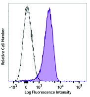
Human Embryonic Kidney 293 (HEK293) cells transfected with human TMEM119* (filled histogram) or transfected with an irrelevant plasmid (open histogram) were stained with 0.6 µg/mL of TMEM119 (Clone A16075D), followed by anti-mouse IgG PE. -

Western blot of anti-TMEM119 (Extracellular) antibody (clone A16075D). Lane 1: Molecular weight marker; Lane 2: Mock transfected HEK293 cell lysate; Lane 3: Lysate of HEK293 cells transfected with human TMEM119*. The blot was incubated with 0.5 µg/mL of the primary antibody overnight at 4°C, followed by incubation with HRP-labeled goat anti-mouse IgG (Cat. No. 405306). β-actin was used as the loading control (Cat. No. 643807). Enhanced chemiluminescence was used as the detection system. -

ICC staining of anti-TMEM119 (Extracellular) antibody (clone A16075D) on HEK293 cells transfected with human TMEM119*. The cells were fixed with 4% PFA, permeabilized with a buffer containing 0.1% Triton X-100 and 0.25% BSA, and blocked with 2% normal goat serum and 0.02% BSA. The cells were then incubated with 5 µg/mL of the primary antibody for overnight at 4°C, followed by incubation wit 1 µg/mL of Alexa Fluor® 594 goat anti-Mouse IgG (Cat. No. 405326) for one hour at room temperature. Nuclei were counterstained with DAPI. The image was captured with a 60X objective. Scale bar: 20 µm -

IHC staining of purified anti-TMEM119 antibody (clone A16075D) on formalin-fixed paraffin-embedded human tonsil tissue. Following antigen retrieval using 0.1 M Tris-buffered saline with Tween 20 (Cat. No. 925501), the tissue was incubated with 5 µg/mL of the primary antibody overnight at 4°C, followed by incubation with 2.5 µg/mL of Alexa Fluor® 647 goat anti-mouse IgG (yellow) (Cat. No. 405322) for one hour at room temperature. Nuclei were counterstained with DAPI (blue), and the slide was mounted with ProLong™ Gold Antifade Mountant. The image was captured with a 10x objective. Scale bar: 100 µm -

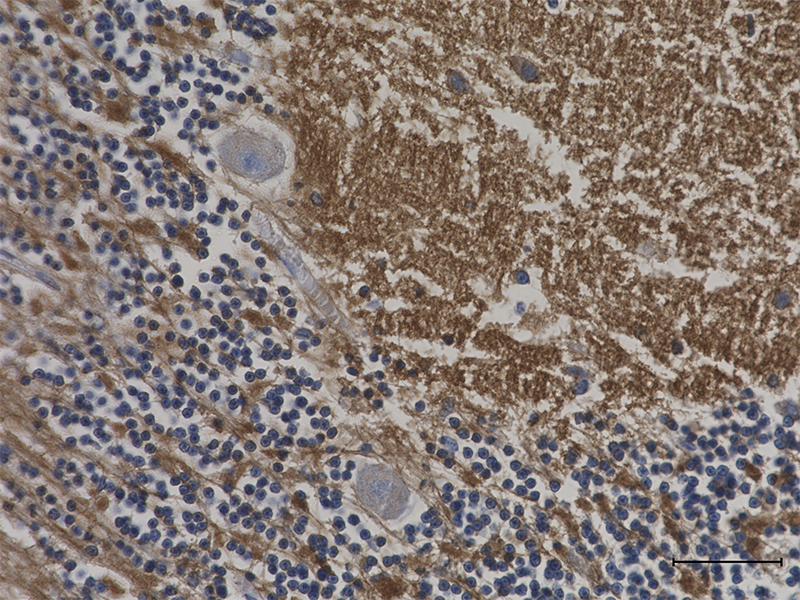
IHC staining of purified anti-TMEM119 antibody (A16075D) on formalin-fixed paraffin-embedded human neocortical brain tissue. Following antigen retrieval using T.E. pH 9.0, the tissue was incubated with 0.5 µg of primary antibody for 1.5 hours at room temperature, followed by incubation with 1:200 dilution of Alexa Fluor® 647 Goat anti-mouse IgG (minimal x-reactivity) antibody (Cat. No. 405322) for 1 hour at room temperature. Nuclei were counterstained with DAPI (Cat. No. 422801). The image was captured with a 40X objective. Scale bar: 50 µm.
| Cat # | Size | Price | Save |
|---|---|---|---|
| 853301 | 25 µg | ¥24,640 | |
| 853302 | 100 µg | ¥63,360 |
Microglia are the resident parenchymal myeloid cells of the CNS, and play an important role in development, homeostasis, disease, and injury. TMEM119 is a transmembrane protein and was identified as a microglia-specific marker in both mouse and human CNS. Moreover, TMEM119 immunoreactivity has been exclusively detected on a subset of Iba1(+) CD68(+) microglia with ramified and amoeboid morphologies in the brains of neurodegenerative diseases. TMEM119 also plays an important role in bone formation and normal bone mineralization. It promotes the differentiation of myoblasts into osteoblasts. and it may induce the commitment and differentiation of myoblasts into osteoblasts. TMEM119 is also essential for normal spermatogenesis and late testicular differentiation.
Product DetailsProduct Details
- Verified Reactivity
- Human
- Antibody Type
- Monoclonal
- Host Species
- Mouse
- Immunogen
- Recombinant human TMEM119 protein (extracellular domain)
- Formulation
- Phosphate-buffered solution, pH 7.2, containing 0.09% sodium azide.
- Preparation
- The antibody was purified by affinity chromatography.
- Concentration
- 0.5 mg/mL
- Storage & Handling
- The antibody solution should be stored undiluted between 2°C and 8°C.
- Application
-
FC - Quality tested
WB, ICC, IHC-P - Verified - Recommended Usage
-
Each lot of this antibody is quality control tested by immunofluorescent staining with flow cytometric analysis. For flow cytometric staining, the suggested use of this reagent is ≤ 0.6 - 5.0 µg/mL. For Western blotting, the suggested use of this reagent is 1.0 - 5.0 µg per mL. For immunocytochemistry, a concentration range of 2.0 - 5.0 µg/mL is suggested. For immunohistochemistry on formalin-fixed paraffin-embedded tissue sections, a concentration range of 5.0 µg/ml is suggested. It is recommended that the reagent be titrated for optimal performance for each application.
- Application Notes
-
For microscopy, the antibody was found to work best when using fresh frozen human tissues. Communication by Dr. Frederick Christian Bennett at Stanford University, under review in Neuron.
*The human TMEM119 full length plasmid was a courtesy gift from Dr. Ben Barres at Stanford University.
-
Application References
(PubMed link indicates BioLegend citation) -
-
Bennett FC, et al. 2018. Neuron. 98:1-14 PubMed
-
- Product Citations
-
- RRID
-
AB_2734646 (BioLegend Cat. No. 853301)
AB_2734647 (BioLegend Cat. No. 853302)
Antigen Details
- Structure
- TMEM119 is an 283 amino acid protein with a molecular mass of 30 kD.
- Distribution
-
Tissue distribution: TMEM119 is expressed in the brain, bone, and lymphoid tissues.
Cellular distribution: Membrane and endoplasmic reticulum. - Function
- TMEM119 is an important microglia marker and is implicated in neurodegenerative diseases.
- Interaction
- TMEM119 interacts with SMAD1, SMAD5 and RUNX2.
- Biology Area
- Cell Biology, Neuroscience, Neuroscience Cell Markers
- Antigen References
-
- Bennett ML, et al. 2016. Proc Natl Acad Sci U S A. 113
- Satoh J, et al. 2016. Neuropathology. 36:39-49
- Kawao N and Kaji H. 2015. J Cell Biochem. 116:687
- Zrzavy T, et al. 2017. Brain. 140:1900-1913
- Gene ID
- 338773 View all products for this Gene ID
- UniProt
- View information about TMEM119 on UniProt.org

Related FAQs
Other Formats
View All TMEM119 Reagents Request Custom Conjugation| Description | Clone | Applications |
|---|---|---|
| Purified anti-TMEM119 (Extracellular) | A16075D | FC,WB,ICC,IHC-P |
| TotalSeq™-A0445 anti-TMEM119 (Extracellular) | A16075D | PG |
| TotalSeq™-B0445 anti-TMEM119 (Extracellular) | A16075D | PG |
| TotalSeq™-C0445 anti-TMEM119 (Extracellular) | A16075D | PG |
| Alexa Fluor® 594 anti-TMEM119 (Extracellular) | A16075D | IHC-P |
| Alexa Fluor® 647 anti-TMEM119 (Extracellular) | A16075D | ICFC,IHC-P,ICC |
| PE anti-TMEM119 (Extracellular) | A16075D | ICFC |
Customers Also Purchased
Compare Data Across All Formats
This data display is provided for general comparisons between formats.
Your actual data may vary due to variations in samples, target cells, instruments and their settings, staining conditions, and other factors.
If you need assistance with selecting the best format contact our expert technical support team.
-
Purified anti-TMEM119 (Extracellular)

Human Embryonic Kidney 293 (HEK293) cells transfected with h... 
Western blot of anti-TMEM119 (Extracellular) antibody (clone... 
ICC staining of anti-TMEM119 (Extracellular) antibody (clone... 
IHC staining of purified anti-TMEM119 antibody (clone A16075... 
IHC staining of purified anti-TMEM119 antibody (A16075D) on ... -
TotalSeq™-A0445 anti-TMEM119 (Extracellular)
-
TotalSeq™-B0445 anti-TMEM119 (Extracellular)
-
TotalSeq™-C0445 anti-TMEM119 (Extracellular)
-
Alexa Fluor® 594 anti-TMEM119 (Extracellular)

IHC staining of Alexa Flour® 594 anti-TMEM119 (clone A16075D... -
Alexa Fluor® 647 anti-TMEM119 (Extracellular)

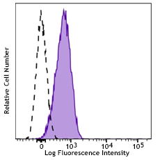
Non-transfected RBL-1 cells (negative control, open histogra... 
IHC staining of Alexa Flour® 647 anti-TMEM119 (clone A16075D... 
Non- transfected RBL-1 cells (negative control, panel A) and... -
PE anti-TMEM119 (Extracellular)

Non-transfected RBL-1 cells (negative control, open histogra...


Follow Us