


- Clone
- D2-40 (See other available formats)
- Regulatory Status
- RUO
- Other Names
- Podoplanin, T1-alpha, hT1alpha-1, hT1alpha-2, PA2.26 antigen, glycoprotein 36, lung type-I cell membrane-associated glycoprotein (T1A-2)
- Isotype
- Mouse IgG1, κ
- Ave. Rating
- Submit a Review
- Product Citations
- publications

-

IHC staining of purified anti-Podoplanin (Lymphatic Endothelial Marker) antibody (clone D2-40) on formalin-fixed paraffin-embedded human colon tissue. The tissue was incubated with 1.3 µg/ml of the primary antibody at room temperature for 60 minutes. BioLegend's Ultra-Streptavidin (USA) HRP kit (Cat. No. 929901) was used for detection followed by hematoxylin counterstaining, according to the protocol provided. The image was captured with a 40X objective. -

IHC staining of purified anti-Podoplanin (Lymphatic Endothelial Marker)antibody (clone D2-40) on formalin-fixed paraffin-embedded human colon tissue. The tissue was incubated with 1 µg/ml of the primary antibody for 60 minutes at room temperature. BioLegend´s Ultra-Streptavidin (USA) HRP kit (Multi-Species, DAB, Cat. No. 929901) was used for detection followed by hematoxylin counterstaining, according to the protocol provided. The image was captured with a 40X objective. -

Western blot analysis of cell lysates from HeLa using Podoplanin Mouse primary antibody and HRP Goat anti-Mouse secondary antibody (Cat. No. 405306). Direct-Blot™ HRP anti-β-actin (Cat. No. 643807) was used as a loading control. -

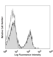
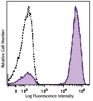
LN319 cells (positive control, filled histogram), or HeLa cells (negative control, open histogram) were fixed and permeabilized using Cyto-Fast™ Fix/Perm Buffer Set (Cat. No. 426803) and stained with Purified anti-Podoplanin (clone D2-40), or Purified mouse IgG1, κ isotype control (open histogram, dashed line) (representative histogram for both cell lines) (Cat. No. 401401), followed by Alexa Fluor® 647 Goat anti-mouse IgG (Cat. No. 405322) -

HeLa cells (negative control, top panels), or LN319 cells (positive control, bottom panels) were fixed and permeabilized with 2% PFA and ice-cold methanol, and then intracellularly stained with Purified anti-Podoplanin (clone D2-40) (right panels) or Purified mouse IgG1, κ isotype control (Cat. No. 401401) (left panels). Nuclei were counterstained with DAPI (Cat. No. 422801) Scale bar 50 µm
| Cat # | Size | Price | Save |
|---|---|---|---|
| 916605 | 25 µg | ¥27,280 | |
| 916606 | 100 µg | ¥58,300 |
Podoplanin is expressed by a variety of normal cells, which include breast and prostate myopepithelial cells, follicular dendritic cells, basal keratinocytes of the skin and cervix (focal), type I pneumocytes, ependymal cells, and fetal cerebral germinal matrix cells. Podoplanin is a marker of lymphatic differentiation because it is expressed by normal human lymphatic endothelium, but not by vascular endothelium. It is diagnostically a useful marker for the evaluation of a variety of neoplasms, including Kaposi sarcoma mesothelioma, testicular germ cell tumors, and cutanneous sebaceous neoplasms.
Product DetailsProduct Details
- Verified Reactivity
- Human
- Antibody Type
- Monoclonal
- Host Species
- Mouse
- Immunogen
- M2A protein derived from human germ cell tumors.
- Formulation
- Phosphate-buffered solution, pH 7.2.
- Preparation
- The antibody was purified by affinity chromatography.
- Concentration
- 1.0 mg/mL
- Storage & Handling
- The antibody solution should be stored undiluted between 2°C and 8°C.
- Application
-
IHC-P - Quality tested
WB, ICFC, ICC - Verified
ELISA Detection - Reported in the literature, not verified in house - Recommended Usage
-
Each lot of this antibody is quality control tested by formalin-fixed paraffin-embedded immunohistochemical staining. For immunohistochemistry, a concentration range of 0.5 - 5.0 μg/mL is suggested. For Western blotting, the suggested use of this reagent is 2.0 - 5.0 µg per mL. For intracellular flow cytometric staining, the suggested use of this reagent is ≤ 0.03 µg per million cells in 100 µL volume. For immunocytochemistry, a concentration range of 1.25 - 5 μg/mL is recommended. It is recommended that the reagent be titrated for optimal performance for each application.
- Application Notes
-
Additional reported applications (for the relevant formats) include: ELISA Detection5 and immunocytochemistry4.
-
Application References
(PubMed link indicates BioLegend citation) -
- Hamanaka T, et al. 2011. Invest. Ophthalmol. Vis. Sci. 52:8849. (IHC-P)
- Choi WW, et al. 2005. Mod. Pathol. 18:143. (IHC-P)
- Chu AY, et al. 2005. Mod. Pathol. 18:105. (IHC-P) PubMed
- Yoon C, et al. 2008. Blood 112:1129. (ICC) PubMed
- Schacht V, et al. 2005. American Journal of Pathology 166:913. (ELISA Detection, WB) PubMed
- Marks A, et al. 1999. British Journal of Cancer 80:569.
- Product Citations
-
- RRID
-
AB_2565819 (BioLegend Cat. No. 916605)
AB_2565820 (BioLegend Cat. No. 916606)
Antigen Details
- Structure
- 35 kD.
- Distribution
-
Membrane and plasma membrane.
- Function
- May be involved in cell migration and/or actin cytoskeleton organization. When expressed in keratinocytes, induces changes in cell morphology with transfected cells showing an elongated shape, numerous membrane protusions, major reorganization of the actin cytoskeleton, increased motility and decreased cell adhesion. Required for normal lung cell proliferation and alveolus formation at birth. Induces platelet aggregation. Does not have any effect on folic acid or amino acid transport and does not function as a water channel or as a regulator of aquaporin-type water channels.
- Interaction
- Cytoskeletal signaling.
- Biology Area
- Cancer Biomarkers, Cell Biology, Neuroinflammation, Neuroscience, Neuroscience Cell Markers
- Gene ID
- 10630 View all products for this Gene ID
- UniProt
- View information about Podoplanin on UniProt.org
Related Pages & Pathways
Pathways


Related FAQs
Other Formats
View All Podoplanin Reagents Request Custom Conjugation| Description | Clone | Applications |
|---|---|---|
| D2-40 Lymphatic Endothelial Marker Monoclonal Antibody | D2-40 | IHC |
| Purified anti-Podoplanin (Lymphatic Endothelial Marker) | D2-40 | IHC-P,WB,ICFC,ICC,ELISA Capture |
| Alexa Fluor® 594 anti-Podoplanin/Lymphatic Endothelial Marker | D2-40 | IHC-P |
| Alexa Fluor® 647 anti-Podoplanin (Lymphatic Endothelial Marker) | D2-40 | IHC-P |
Customers Also Purchased
Compare Data Across All Formats
This data display is provided for general comparisons between formats.
Your actual data may vary due to variations in samples, target cells, instruments and their settings, staining conditions, and other factors.
If you need assistance with selecting the best format contact our expert technical support team.
-
D2-40 Lymphatic Endothelial Marker Monoclonal Antibody

Immunohistochemistry showing lymphatic channels with stainin... 

-
Purified anti-Podoplanin (Lymphatic Endothelial Marker)

Western blot analysis of cell lysates from HeLa using Podopl... 
IHC staining of purified anti-Podoplanin (Lymphatic Endothel... 
IHC staining of purified anti-Podoplanin (Lymphatic Endothel... 
LN319 cells (positive control, filled histogram), or HeLa ce... 
HeLa cells (negative control, top panels), or LN319 cells (p... -
Alexa Fluor® 594 anti-Podoplanin/Lymphatic Endothelial Marker

Human paraffin-embedded breast tissue slices were prepared ... -
Alexa Fluor® 647 anti-Podoplanin (Lymphatic Endothelial Marker)

Human paraffin-embedded breast tissue slices were prepared w...


Follow Us