


- Clone
- DU46-1.1 (See other available formats)
- Regulatory Status
- RUO
- Other Names
- PTEN induced putative kinase 1, BRPK, PARK6, Serine/threonine-protein kinase PINK1
- Isotype
- Mouse IgG1, κ
- Ave. Rating
- Submit a Review
- Product Citations
- publications

-

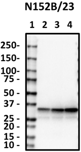
Western blot of purified anti-PINK1 antibody (clone DU46-1.1). Lane 1: Molecular weight marker; Lane 2: 20 µg of control HeLa cell lysate; Lane 3: 20 µg of CCCP-treated HeLa cell lysate. The blot was incubated with 5 µg/mL of the primary antibody overnight at 4°C, followed by incubation with HRP labeled goat anti-mouse IgG (Cat. No. 405306). Enhanced chemiluminescence was used as the detection system. -

ICC staining of purified anti-PINK1 antibody (clone DU46-1.1) on control (left) or CCCP (right) treated HeLa cells. The cells were treated with 10 µM of CCCP for 24 hours. The cells were fixed with 4% PFA, permeabilized with a buffer containing 0.1% Triton X-100 and 0.25% BSA, and blocked with 2% normal goat serum and 0.02% BSA. The cells were then incubated with 5 μg/mL of the primary antibody overnight at 4°C, followed by incubation with 2.5 µg/ml of Alexa Fluor® 594 Goat anti-mouse IgG (Cat. No. 405326) for one hour at room temperature. The cells were counterstained with Flash Phalloidin™ Green 488 (Cat. No. 424201) at a 1:100 dilution for 20min at room temperature. The slides were mounted with fluoromount G with DAPI. The images were captured with a 60X objective. Scale bar: 20 µm
| Cat # | Size | Price | Save |
|---|---|---|---|
| 846201 | 25 µg | ¥23,320 | |
| 846202 | 100 µg | ¥58,300 |
PINK1 (PTEN-induced putative kinase 1) is a 63 kD mitochondrial transmembrane protein that is often cleaved by PARL into a 53 kD fragment. The protein is involved in the clearance of damaged mitochondria via selective autophagy (mitophagy). PINK1 phosphorylation of mitochondrial proteins can protect against mitochondrial dysfunction due to cellular stresses. PINK1 mutations can cause autosomal recessive early-onset Parkinson disease.
Product DetailsProduct Details
- Verified Reactivity
- Human
- Antibody Type
- Monoclonal
- Host Species
- Mouse
- Formulation
- Phosphate-buffered solution, pH 7.2, containing 0.09% sodium azide.
- Preparation
- The antibody was purified by affinity chromatography.
- Concentration
- 0.5 mg/ml
- Storage & Handling
- The antibody solution should be stored undiluted between 2°C and 8°C.
- Application
-
WB - Quality tested
ICC - Verified - Recommended Usage
-
Each lot of this antibody is quality control tested by Western blotting. For Western blotting, the suggested use of this reagent is 5.0 - 10 µg per mL. For immunocytochemistry, a concentration range of 1.0 - 5.0 μg/mL is recommended. It is recommended that the reagent be titrated for optimal performance for each application.
- Product Citations
-
- RRID
-
AB_2783414 (BioLegend Cat. No. 846201)
AB_2572127 (BioLegend Cat. No. 846202)
Antigen Details
- Structure
- PINK1 is a 581 amino acid, 63 kD, transmembrane protein that contains an N-terminal mitochondrial localization sequence, a serine/threonine kinase domain, and a C-terminal regulatory domain. PINK1 is proteolytically cleaved by PARL into a 55 kD fragment.
- Distribution
-
Mitochondria and cytoplasm.
- Function
- PINK1 is involved in the clearance of damaged mitochondria via selective autophagy (mitophagy) by mediating activation and translocation of Parkin protein (PARK2). PARL-cleaved PINK1 is a cytoplasmic probe for damaged mitochondria. PINK1 phosphorylation of mitochondrial proteins can protect against mitochondrial dysfunction due to cellular stresses.
- Interaction
- PARKIN, Beclin 1, FBX07, DJ-1, HSP90/CDC37.
- Ligand/Receptor
- PARKIN, Beclin 1, FBX07, DJ-1, HSP90/CDC37.
- Biology Area
- Cell Biology, Mitochondrial Function, Neurodegeneration, Neuroinflammation, Neuroscience, Neuroscience Cell Markers, Protein Trafficking and Clearance
- Molecular Family
- Mitochondrial Markers
- Antigen References
-
1. Trempe JF, Fon EA. 2013. Front Neurol. 4:38.
2. Durcan TM, Fon EA. 2015. Genes Dev. 29(10):989. - Gene ID
- 65018 View all products for this Gene ID
- UniProt
- View information about PINK1 on UniProt.org

Related FAQs
Other Formats
View All PINK1 Reagents Request Custom Conjugation| Description | Clone | Applications |
|---|---|---|
| Purified anti-PINK1 | DU46-1.1 | WB,ICC |
Customers Also Purchased
Compare Data Across All Formats
This data display is provided for general comparisons between formats.
Your actual data may vary due to variations in samples, target cells, instruments and their settings, staining conditions, and other factors.
If you need assistance with selecting the best format contact our expert technical support team.


Follow Us