


- Clone
- 93 (See other available formats)
- Regulatory Status
- RUO
- Other Names
- Fcγ R III/II, Ly-17
- Isotype
- Rat IgG2a, λ
- Ave. Rating
- Submit a Review
- Product Citations
- publications

-

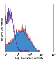
C57BL/6 mouse splenocytes stained with 93 PE
| Cat # | Size | Price | Save |
|---|---|---|---|
| 101307 | 50 µg | ¥21,560 | |
| 101308 | 200 µg | ¥66,880 |
CD16 is low affinity IgG Fc receptor III (FcR III) and CD32 is FcR II. CD16/CD32 are expressed on B cells, monocytes/macrophages, NK cells, granulocytes, mast cells, and dendritic cells. The Fc receptors bind antibody-antigen immune complexes and mediate adaptive immune responses.
Product DetailsProduct Details
- Verified Reactivity
- Mouse
- Antibody Type
- Monoclonal
- Host Species
- Rat
- Immunogen
- Sorted pre-B cells
- Formulation
- Phosphate-buffered solution, pH 7.2, containing 0.09% sodium azide.
- Preparation
- The antibody was purified by affinity chromatography, and conjugated with PE under optimal conditions.
- Concentration
- 0.2 mg/ml
- Storage & Handling
- The antibody solution should be stored undiluted between 2°C and 8°C, and protected from prolonged exposure to light. Do not freeze.
- Application
-
FC - Quality tested
- Recommended Usage
-
Each lot of this antibody is quality control tested by immunofluorescent staining with flow cytometric analysis. For flow cytometric staining, the suggested use of this reagent is ≤1.0 µg per million cells in 100 µl volume. It is recommended that the reagent be titrated for optimal performance for each application.
- Excitation Laser
-
Blue Laser (488 nm)
Green Laser (532 nm)/Yellow-Green Laser (561 nm)
- Application Notes
-
Clone 93 can be used for blocking of CD16/CD32 interactions with the Fc domain of immunoglobulins, but is not the same clone as 2.4G2.
The 93 mAb is specific to the common epitope of CD16/CD32. Additional reported applications (for the relevant formats) include: immunoprecipitation1 and blocking of Fc-mediated reactions in functional studies2,4,23. It is useful for blocking non-specific binding of immunoglobulin to Fc receptors. For blocking of Fc receptors in flow cytometric analysis, pre-incubate the cells with purified anti-CD16/CD32 antibody (=1.0 µg per 106 cells in 100 µL volume) for 5-10 minutes on ice prior to immunostaining. For highly sensitive assays, we recommend Ultra-LEAF™ purified antibody (Cat. No. 101330) (Endotoxin <0.01 EU/µg, Azide-Free, 0.2 µm filtered). -
Application References
(PubMed link indicates BioLegend citation) -
- Personal communication (IP)
- Oliver AM, et al. 1999. Hybridoma 18:113. (Block)
- Brummel R and Lenert P. 2005. J. Immunol. 174:2429.
- Terrazas LI, et al. 2005. Int. J. Parasitol. 35:1349. (Block)
- Clements JL, et al. 2006. J. Immunol. 177:905.
- Mohamed W, et al. 2010. Infect Immun. 78:3306. PubMed
- Ouchi T, et al. 2011. J. Exp Med. 208:2607. PubMed
- Kmieciak M, et al. 2011. J. Vis. Exp. 47:2381. PubMed
- Yamazaki S, et al. 2012. PLoS One. 7:e51665. PubMed
- Li J, et al. 2012. Arthritis Rheum. 64:1098. PubMed
- Azuma M, et al. 2012. Oncoimmunology. 1:581. PubMed
- Koon HW, et al. 2013. J. Vis. Exp. 68:4208. PubMed
- Hegde VL, et al. 2013. J Biol Chem. 288:36810. PubMed
- Huang J, et al. 2013. J. Immunol Methods. 387:254. PubMed
- Dutow P, et al. 2014. J Infect Dis. PubMed
- Fan Y, et al. 2014. J Exp Med. 211:313. PubMed
- Huang HN, et al. 2014. Antimicrob Agents Chemother. 58:1538. PubMed
- Takei S, et al. 2014. Vaccine. 32:3066. PubMed
- Richardson ML, et al. 2014. PLoS Negl Trop Dis. 8:2825. PubMed
- Cekanaviciute E, et al. 2014. J Immunol. 193:139. PubMed
- Kimura T, et al. 2014. Int Immunol. 26:697. PubMed
- Everad A, et al. 2014. Nat Commun. 5:5648. PubMed
- Cenci E, et al. 2006. J. Leuko. Biol. 79(1):40-5. (Block)
- Product Citations
-
- RRID
-
AB_312806 (BioLegend Cat. No. 101307)
AB_312807 (BioLegend Cat. No. 101308)
Antigen Details
- Structure
- Ig superfamily, 40-60 kD
- Distribution
-
B cells, monocyte/macrophages, NK cells, neutrophils, mast cells, dendritic cells
- Function
- Low affinity receptors for IgG
- Ligand/Receptor
- IgG
- Cell Type
- B cells, Dendritic cells, Macrophages, Mast cells, Monocytes, Neutrophils, NK cells
- Biology Area
- Immunology, Innate Immunity
- Molecular Family
- CD Molecules, Fc Receptors
- Antigen References
-
1. Barclay AN, et al. 1997. The Leukocyte Antigen FactsBook Academic Press.
2. Unkeless JC. 1989. J. Clin. Invest. 83:355.
3. Qiu WQ, et al. 1990. Science 248:732. - Gene ID
- 14130 View all products for this Gene ID 14131 View all products for this Gene ID
- UniProt
- View information about CD16/32 on UniProt.org


Related FAQs
- What type of PE do you use in your conjugates?
- We use R-PE in our conjugates.
Other Formats
View All CD16/32 Reagents Request Custom Conjugation| Description | Clone | Applications |
|---|---|---|
| Biotin anti-mouse CD16/32 | 93 | FC |
| FITC anti-mouse CD16/32 | 93 | FC |
| PE anti-mouse CD16/32 | 93 | FC |
| Purified anti-mouse CD16/32 | 93 | FC,IP,Block |
| Ultra-LEAF™ Purified anti-mouse CD16/32 | 93 | FC,IP,Block |
| Alexa Fluor® 647 anti-mouse CD16/32 | 93 | FC |
| PE/Cyanine7 anti-mouse CD16/32 | 93 | FC |
| TruStain FcX™ (anti-mouse CD16/32) | 93 | FC |
| PerCP/Cyanine5.5 anti-mouse CD16/32 | 93 | FC |
| APC anti-mouse CD16/32 | 93 | FC |
| APC/Cyanine7 anti-mouse CD16/32 | 93 | FC |
| Brilliant Violet 421™ anti-mouse CD16/32 | 93 | FC |
| Brilliant Violet 510™ anti-mouse CD16/32 | 93 | FC |
| Purified anti-mouse CD16/32 (Maxpar® Ready) | 93 | FC,CyTOF® |
| Brilliant Violet 711™ anti-mouse CD16/32 | 93 | FC |
| TotalSeq™-A0109 anti-mouse CD16/32 | 93 | PG |
| TotalSeq™-B0109 anti-mouse CD16/32 | 93 | PG |
| TotalSeq™-C0109 anti-mouse CD16/32 | 93 | PG |
Customers Also Purchased
Compare Data Across All Formats
This data display is provided for general comparisons between formats.
Your actual data may vary due to variations in samples, target cells, instruments and their settings, staining conditions, and other factors.
If you need assistance with selecting the best format contact our expert technical support team.
-
Biotin anti-mouse CD16/32

C57BL/6 mouse splenocytes stained with biotinylated 93, foll... -
FITC anti-mouse CD16/32

C57BL/6 mouse splenocytes stained with 93 FITC -
PE anti-mouse CD16/32

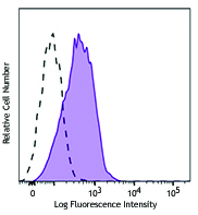
C57BL/6 mouse splenocytes stained with 93 PE -
Purified anti-mouse CD16/32

C57BL/6 mouse splenocytes stained with purified 93, followed... -
Ultra-LEAF™ Purified anti-mouse CD16/32

C57BL/6 mouse splenocytes stained with Ultra-LEAF™ purified ... -
Alexa Fluor® 647 anti-mouse CD16/32

C57BL/6 mouse splenocytes stained with Alexa Fluor® 647 -
PE/Cyanine7 anti-mouse CD16/32

C57BL/6 splenocytes stained with 93 PE/Cyanine7 -
TruStain FcX™ (anti-mouse CD16/32)
-
PerCP/Cyanine5.5 anti-mouse CD16/32

C57BL/6 mouse splenocytes stained with CD16/32 (clone 93) Pe... -
APC anti-mouse CD16/32

C57BL/6 splenocytes stained with 93 APC -
APC/Cyanine7 anti-mouse CD16/32

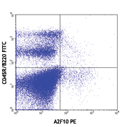
BALB/c splenocytes stained with CD45R/B220 FITC and CD16/32 ... -
Brilliant Violet 421™ anti-mouse CD16/32

C57BL/6 mouse splenocytes were stained with CD16/32 (clone 9... -
Brilliant Violet 510™ anti-mouse CD16/32

C57BL/6 mouse splenocytes were stained with CD16/32 (clone 9... -
Purified anti-mouse CD16/32 (Maxpar® Ready)

C57BL/6 mouse splenocytes stained with 148Nd-anti-CD11b (M1/... -
Brilliant Violet 711™ anti-mouse CD16/32

C57BL/6 mouse splenocytes were stained with CD16/32 (clone 9... -
TotalSeq™-A0109 anti-mouse CD16/32
-
TotalSeq™-B0109 anti-mouse CD16/32
-
TotalSeq™-C0109 anti-mouse CD16/32


Follow Us