


- Clone
- SK3 (See other available formats)
- Regulatory Status
- RUO
- Other Names
- T4, Leu3a
- Isotype
- Mouse IgG1, κ
- Ave. Rating
- Submit a Review
- Product Citations
- publications

-

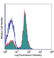
Human peripheral blood lymphocytes were stained with CD4 (clone SK3) Brilliant Violet 750™ (filled histogram) or mouse IgG1, κ Brilliant Violet 750™ isotype control (open histogram).
| Cat # | Size | Price | Save |
|---|---|---|---|
| 344643 | 25 tests | ¥45,980 | |
| 344644 | 100 tests | ¥91,960 |
CD4, also known as T4, is a 55 kD single-chain type I transmembrane glycoprotein expressed on most thymocytes, a subset of T cells, and monocytes/macrophages. CD4, a member of the Ig superfamily, recognizes antigens associated with MHC class II molecules and participates in cell-cell interactions, thymic differentiation, and signal transduction. CD4 acts as a primary receptor for HIV, binding to HIV gp120. CD4 has also been shown to interact with IL-16.
Product DetailsProduct Details
- Verified Reactivity
- Human
- Antibody Type
- Monoclonal
- Host Species
- Mouse
- Formulation
- Phosphate-buffered solution, pH 7.2, containing 0.09% sodium azide and BSA (origin USA).
- Preparation
- The antibody was purified by affinity chromatography and conjugated with Brilliant Violet 750™ under optimal conditions.
- Concentration
- Lot-specific (to obtain lot-specific concentration and expiration, please enter the lot number in our Certificate of Analysis online tool.)
- Storage & Handling
- The antibody solution should be stored undiluted between 2°C and 8°C, and protected from prolonged exposure to light. Do not freeze.
- Application
-
FC - Quality tested
- Recommended Usage
-
Each lot of this antibody is quality control tested by immunofluorescent staining with flow cytometric analysis. For flow cytometric staining, the suggested use of this reagent is 5 µl per million cells in 100 µl staining volume or 5 µl per 100 µl of whole blood.
Brilliant Violet 750™ excites at 405 nm and emits at 750 nm. The bandpass filter 780/60 nm is recommended for detection, although filter optimization may be required depending on other fluorophores used. Be sure to verify that your cytometer configuration and software setup are appropriate for detecting this channel. Refer to your instrument manual or manufacturer for support. Brilliant Violet 750™ is a trademark of Sirigen Group Ltd.
Learn more about Brilliant Violet™.
This product is subject to proprietary rights of Sirigen Inc. and is made and sold under license from Sirigen Inc. The purchase of this product conveys to the buyer a non-transferable right to use the purchased product for research purposes only. This product may not be resold or incorporated in any manner into another product for resale. Any use for therapeutics or diagnostics is strictly prohibited. This product is covered by U.S. Patent(s), pending patent applications and foreign equivalents. - Excitation Laser
-
Violet Laser (405 nm)
-
Application References
(PubMed link indicates BioLegend citation) -
- Evans RL, et al. 1981. Immunol. 78:544
- Arno A et al. 1999. J. Infect. Dis. 180:56
- Muech M, et al. 1997. Blood 89:1364
- Wang L, et al. 2012. Cytometry A. 81:567. PubMed
- RRID
-
AB_2734346 (BioLegend Cat. No. 344643)
AB_2800920 (BioLegend Cat. No. 344644)
Antigen Details
- Structure
- Ig superfamily, type I transmembrane glycoprotein, 55 kD
- Distribution
-
T cell subset, majority of thymocytes, monocytes/macrophages
- Function
- MHC class II co-receptor, lymphocyte adhesion, thymic differentiation, HIV receptor
- Ligand/Receptor
- MHC class II molecules, HIV gp120, IL-16
- Cross
- T cell subset, majority of thymocytes, monocytes/macrophages
- Cell Type
- Macrophages, Monocytes, T cells, Thymocytes, Tregs
- Biology Area
- Immunology
- Molecular Family
- CD Molecules
- Antigen References
-
1. Center D et al. 1996. Immunol. Today 17:476.
2. Gaubin M et al. 1996. Eur. J. Clin. Chem. Clin. Biochem. 34:723. - Gene ID
- 920 View all products for this Gene ID
- UniProt
- View information about CD4 on UniProt.org
Related Pages & Pathways
Pathways


Related FAQs
Other Formats
View All CD4 Reagents Request Custom ConjugationCustomers Also Purchased
Compare Data Across All Formats
This data display is provided for general comparisons between formats.
Your actual data may vary due to variations in samples, target cells, instruments and their settings, staining conditions, and other factors.
If you need assistance with selecting the best format contact our expert technical support team.
-
PerCP anti-human CD4

Human peripheral blood lymphocytes were stained with CD4 (cl... -
Purified anti-human CD4

Human peripheral blood lymphocytes stained with SK3, followe... -
FITC anti-human CD4

Human peripheral blood lymphocytes stained with SK3 FITC -
PE anti-human CD4

Human peripheral blood lymphocytes stained with SK3 PE -
PerCP/Cyanine5.5 anti-human CD4

Human peripheral blood lymphocytes were stained with anti-hu... -
Biotin anti-human CD4

Human peripheral blood lymphocytes stained with biotinylated... -
PE/Cyanine7 anti-human CD4

Human peripheral blood lymphocytes stained with SK3 PE/Cyani... -
APC anti-human CD4

Human peripheral blood lymphocytes stained with SK3 APC -
APC/Cyanine7 anti-human CD4

Human peripheral blood lymphocytes were stained with anti-hu... -
Alexa Fluor® 488 anti-human CD4

Human peripheral blood lymphocytes stained with SK3 Alexa Fl... -
Pacific Blue™ anti-human CD4

Human peripheral blood lymphocytes stained with SK3 Pacific ... -
Alexa Fluor® 700 anti-human CD4

Human peripheral blood lymphocytes were stained with CD4 (cl... -
Purified anti-human CD4 (Maxpar® Ready)

Human PBMCs stained with 154Sm-anti-CD45 (HI30) and 174Yb-an... -
PE anti-human CD4

Typical results from human peripheral blood lymphocytes stai... -
Brilliant Violet 421™ anti-human CD4

Human peripheral blood lymphocytes were stained with CD4 (cl... -
Brilliant Violet 510™ anti-human CD4

Human peripheral blood lymphocytes were stained with CD4 (cl... -
Alexa Fluor® 647 anti-human CD4

Human peripheral blood lymphocytes were stained with CD4 (cl... -
FITC anti-human CD4

Typical results from human peripheral blood lymphocytes stai... -
APC/Fire™ 750 anti-human CD4

Human peripheral blood lymphocytes were stained with CD3 PE ... 
-
PE/Dazzle™ 594 anti-human CD4

Human peripheral blood lymphocytes were stained with CD4 (cl... -
Pacific Blue™ anti-human CD4

Typical results from human peripheral blood lymphocytes stai... -
Brilliant Violet 785™ anti-human CD4

Human peripheral blood lymphocytes were stained with CD4 (cl... -
Brilliant Violet 750™ anti-human CD4

Human peripheral blood lymphocytes were stained with CD4 (cl... -
Brilliant Violet 605™ anti-human CD4

Human peripheral blood lymphocytes were stained with CD4 (cl... -
Brilliant Violet 711™ anti-human CD4

Human peripheral blood lymphocytes were stained with CD4 (cl... -
PE/Cyanine7 anti-human CD4
Typical results from human peripheral blood lymphocytes sta... -
TotalSeq™-A0045 anti-human CD4
-
TotalSeq™-C0045 anti-human CD4
-
PE/Cyanine5 anti-human CD4

Human peripheral blood lymphocytes were stained with anti-hu... -
PerCP/Cyanine5.5 anti-human CD4

Typical results from human peripheral blood lymphocytes stai... -
Spark Blue™ 550 anti-human CD4

Human peripheral blood lymphocytes were stained with CD4 (SK... -
Spark NIR™ 685 anti-human CD4

Human peripheral blood lymphocytes were stained with CD3 (cl... -
APC anti-human CD4
Typical results from human peripheral blood lymphocytes stai... -
KIRAVIA Blue 520™ anti-human CD4

Human peripheral blood lymphocytes were stained with CD3 APC... -
APC/Fire™ 810 anti-human CD4

Human peripheral blood lymphocytes were stained with CD4 (cl... -
PE/Fire™ 640 anti-human CD4

Human peripheral blood lymphocytes were stained with CD4 (cl... -
PE/Fire™ 700 anti-human CD4

Human peripheral blood lymphocytes were stained with anti-hu... -
PerCP anti-human CD4

Typical results from human peripheral blood lymphocytes stai... -
Spark Violet™ 538 anti-human CD4 Antibody

Human peripheral blood lymphocytes were stained with CD4 (cl... -
Spark YG™ 581 anti-human CD4

Human peripheral blood lymphocytes were stained with anti-hu... -
Alexa Fluor® 660 anti-human CD4

Human peripheral blood lymphocytes were stained with CD4 (cl... -
Spark YG™ 593 anti-human CD4

Human peripheral blood lymphocytes were stained with anti-hu... -
PE/Fire™ 810 anti-human CD4 Antibody

Human peripheral blood lymphocytes were stained with anti-hu... -
APC/Fire™ 750 anti-human CD4

Typical results from human peripheral blood lymphocytes stai... -
Spark Blue™ 574 anti-human CD4

Human peripheral lymphocytes were stained with anti-human CD... -
GMP APC anti-human CD4

Typical results from human peripheral blood lymphocytes stai... -
TotalSeq™-B0045 anti-human CD4 Antibody
-
Spark Violet™ 423 anti-human CD4 Antibody

Human peripheral blood lymphocytes were stained with anti-hu... -
GMP Pacific Blue™ anti-human CD4

Typical results from human peripheral blood lymphocytes stai... -
Spark UV™ 387 anti-human CD4

Human peripheral blood lymphocytes were stained with anti-hu... -
GMP PE anti-human CD4

Typical results from human peripheral blood lymphocytes stai... -
GMP FITC anti-human CD4

Typical results from human peripheral blood lymphocytes stai... -
Spark Red™ 718 anti-human CD4

Human peripheral blood lymphocytes were stained with anti-hu... -
GMP PerCP anti-human CD4

Typical results from human peripheral blood lymphocytes stai... -
GMP PE/Cyanine7 anti-human CD4

Typical results from human peripheral blood lymphocytes stai... -
Spark Violet™ 500 anti-human CD4

Human peripheral blood lymphocytes were stained with anti-hu... -
GMP PerCP/Cyanine5.5 anti-human CD4

Typical results from human peripheral blood lymphocytes stai... -
Brilliant Violet 650™ anti-human CD4

Human peripheral blood lymphocytes were stained with anti-hu... -
PerCP/Fire™ 806 anti-human CD4

Human peripheral blood lymphocytes were stained with anti-hu... -
Spark Blue™ 515 anti-human CD4

Human peripheral blood lymphocytes were stained with anti-hu... -
GMP APC/Fire™ 750 anti-human CD4

Typical results from human peripheral blood lymphocytes stai... -
Spark Violet™ 423 anti-human CD4

Typical results from human peripheral blood lymphocytes stai... -
PerCP/Fire™ 780 anti-human CD4

Human peripheral blood lymphocytes were stained with anti-hu... -
PE/Fire™ 744 anti-human CD4

Human peripheral blood mononuclear cells were stained with a... -
Spark PLUS UV™ 395 anti-human CD4

Human peripheral blood lymphocytes were stained anti-human C...


Follow Us