


- Clone
- M80 (See other available formats)
- Regulatory Status
- RUO
- Other Names
- Thrombomodulin, TM, THRM, THBD, Fetomodulin, BDCA-3
- Isotype
- Mouse IgG1, κ
- Ave. Rating
- Submit a Review
- Product Citations
- publications

-

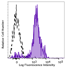
LPS-stimulated (overnight) human peripheral blood monocytes were stained with CD141 (clone M80) Brilliant Violet 605™ (filled histogram) or mouse IgG1, κ Brilliant Violet 605™ isotype control (open histogram). Data shown was gated on monocyte population.
| Cat # | Size | Price | Save |
|---|---|---|---|
| 344117 | 25 tests | ¥47,300 | |
| 344118 | 100 tests | ¥98,120 |
CD141 is a 75 kD, single chain, type I membrane glycoprotein also known as thrombomodulin, TM, THRM, THBD, and fetomodulin. CD141 is an important cofactor in the protein C anticoagulant system. After binding to its ligand thrombin, CD141 activates protein C, which degrades clotting factors Va and VIIIa, and as a consequence the amount of thrombin is reduced. CD141 is expressed on macrophages, monocytes, a subpopulation of myeloid dendritic cells, vascular endothelial cells, and keratinocytes. Besides anti-coagulation function, CD141 is also involved in embryonic and atherosclerotic plaque development.
Product DetailsProduct Details
- Verified Reactivity
- Human
- Reported Reactivity
- African Green, Baboon
- Antibody Type
- Monoclonal
- Host Species
- Mouse
- Formulation
- Phosphate-buffered solution, pH 7.2, containing 0.09% sodium azide and BSA (origin USA).
- Preparation
- The antibody was purified by affinity chromatography and conjugated with Brilliant Violet 605™ under optimal conditions.
- Concentration
- Lot-specific (to obtain lot-specific concentration and expiration, please enter the lot number in our Certificate of Analysis online tool.)
- Storage & Handling
- The antibody solution should be stored undiluted between 2°C and 8°C, and protected from prolonged exposure to light. Do not freeze.
- Application
-
FC - Quality tested
- Recommended Usage
-
Each lot of this antibody is quality control tested by immunofluorescent staining with flow cytometric analysis. For flow cytometric staining, the suggested use of this reagent is 5 µl per million cells in 100 µl staining volume or 5 µl per 100 µl of whole blood.
Brilliant Violet 605™ excites at 405 nm and emits at 603 nm. The bandpass filter 610/20 nm is recommended for detection, although filter optimization may be required depending on other fluorophores used. Be sure to verify that your cytometer configuration and software setup are appropriate for detecting this channel. Refer to your instrument manual or manufacturer for support. Brilliant Violet 605™ is a trademark of Sirigen Group Ltd.
Learn more about Brilliant Violet™.
This product is subject to proprietary rights of Sirigen Inc. and is made and sold under license from Sirigen Inc. The purchase of this product conveys to the buyer a non-transferable right to use the purchased product for research purposes only. This product may not be resold or incorporated in any manner into another product for resale. Any use for therapeutics or diagnostics is strictly prohibited. This product is covered by U.S. Patent(s), pending patent applications and foreign equivalents. - Excitation Laser
-
Violet Laser (405 nm)
- Product Citations
-
- RRID
-
AB_2572198 (BioLegend Cat. No. 344117)
AB_2572199 (BioLegend Cat. No. 344118)
Antigen Details
- Structure
- Single chain, type I membrane glycoprotein, 75 kD
- Distribution
-
Macrophages, monocytes, subset of myeloid dendritic cells, vascular endothelial cells, keratinocytes
- Function
- After thrombin binding, CD141 activates protein C, which degrades clotting factors Va and VIIIa and reduces the amount of thrombin generated.
- Interaction
- Protein C, Thrombin-activatable fibrinolysis inhibitor (TAFI), Platelet factor 4 (PF4)
- Ligand/Receptor
- Thrombin
- Cell Type
- Dendritic cells, Endothelial cells, Macrophages, Monocytes
- Biology Area
- Immunology, Innate Immunity
- Molecular Family
- CD Molecules
- Antigen References
-
1. Suzuki K, et al. 1987. EMBO J. 6:1891.
2. Esmon CT, et al. 1989. J. Biol. Chem. 264:4743.
3. Delvaeye M, et al. 2009. N. Engl. J. Med. 361:345.
4. Shi CS, et al. 2008. Blood 112:3661.
5. Chen LC, et al. 2009. J. Infect. 58:368. - Gene ID
- 7056 View all products for this Gene ID
- UniProt
- View information about CD141 on UniProt.org


Related FAQs
Other Formats
View All CD141 Reagents Request Custom ConjugationCustomers Also Purchased
Compare Data Across All Formats
This data display is provided for general comparisons between formats.
Your actual data may vary due to variations in samples, target cells, instruments and their settings, staining conditions, and other factors.
If you need assistance with selecting the best format contact our expert technical support team.
-
Purified anti-human CD141 (Thrombomodulin)

Human peripheral blood monocytes stained with M80 PE 
LPS-stimulated (overnight) human peripheral blood mononuclea... -
PE anti-human CD141 (Thrombomodulin)
Human peripheral blood monocytes stained with M80 PE
LPS-stimulated (overnight) human peripheral blood mononuclea... -
APC anti-human CD141 (Thrombomodulin)

Human peripheral blood monocytes were stimulated with LPS (o... -
Biotin anti-human CD141 (Thrombomodulin)

Human peripheral blood monocytes were stained with biotinyla... -
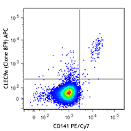
PE/Cyanine7 anti-human CD141 (Thrombomodulin)

LPS-stimulated (overnight) human peripheral blood monocytes ... -
PerCP/Cyanine5.5 anti-human CD141 (Thrombomodulin)

LPS-stimulated (overnight) human peripheral blood monocytes ... -
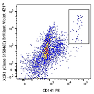
Brilliant Violet 421™ anti-human CD141 (Thrombomodulin)

LPS-stimulated (overnight) human peripheral blood monocytes ... -
Brilliant Violet 785™ anti-human CD141 (Thrombomodulin)

LPS-stimulated (overnight) human peripheral blood monocytes ... -
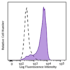
Brilliant Violet 605™ anti-human CD141 (Thrombomodulin)

LPS-stimulated (overnight) human peripheral blood monocytes ... -
PE/Dazzle™ 594 anti-human CD141 (Thrombomodulin)

Human peripheral blood monocytes were stimulated with LPS (o... -
TotalSeq™-A0163 anti-human CD141 (Thrombomodulin)
-
Alexa Fluor® 647 anti-human CD141 (Thrombomodulin)

Human peripheral blood monocytes were stained with CD141 (Th... -
TotalSeq™-C0163 anti-human CD141 (Thrombomodulin)
-
TotalSeq™-B0163 anti-human CD141 (Thrombomodulin)
-
KIRAVIA Blue 520™ anti-human CD141 (Thrombomodulin)

Human peripheral blood monocytes were stained with anti-huma... -
TotalSeq™-D0163 anti-human CD141 (Thrombomodulin)
-
PE/Fire™ 810 anti-human CD141 (Thrombomodulin)

Human peripheral blood monocytes were stained with anti-huma... -
Brilliant Violet 711™ anti-human CD141 (Thrombomodulin)

Human peripheral blood monocytes were stained with anti-huma...


Follow Us