


- Clone
- BM8 (See other available formats)
- Regulatory Status
- RUO
- Other Names
- EMR1, Ly71
- Isotype
- Rat IgG2a, κ
- Ave. Rating
- Submit a Review
- Product Citations
- publications

-

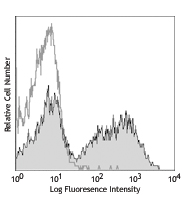
Thioglycolate-elicited Balb/c mouse peritoneal macrophages were stained with F4/80 (clone BM8) Brilliant Violet 421™ (filled histogram) or rat IgG2a, κ Brilliant Violet 421™ isotype control (open histogram). -

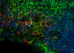
C57BL/6 mouse frozen spleen section was fixed with 4% paraformaldehyde (PFA) for ten minutes at room temperature and blocked with 5% FBS plus 5% rat/mouse serum for 30 minutes at room temperature. Then the section was stained with 2 µg/ml of anti-mouse F4/80 (clone BM8) Brilliant Violet 421™ (Blue) and anti-mouse CD8a (clone 53-6.7) Alexa Fluor® 647 (red) overnight at 4°C. The image was captured with a 10X objective. -

Confocal image of C57BL/6 mouse spleen sample acquired using the IBEX method of highly multiplexed antibody-based imaging: MHCII (blue) in Cycle 2 and F4/80 (magenta) in Cycle 2. Tissues were prepared using ~1% (vol/vol) formaldehyde and a detergent. Following fixation, samples are immersed in 30% (wt/vol) sucrose for cryoprotection. Images are courtesy of Drs. Andrea J. Radtke and Ronald N. Germain of the Center for Advanced Tissue Imaging (CAT-I) in the National Institute of Allergy and Infectious Diseases (NIAID, NIH). -

Mice were injected subcutaneously with sheep red blood cells in a volume of 25 µl per site on days 0 and 4 and harvested on day 11. Confocal image of C57BL/6 mouse lymph node acquired using the IBEX method of highly multiplexed antibody-based imaging: F4/80 (cyan) in Cycle 3, CD68 (blue) in Cycle 6, and NK1.1 (magenta) in Cycle 9. Tissues were prepared using ~1% (vol/vol) formaldehyde and a detergent. Following fixation, samples are immersed in 30% (wt/vol) sucrose for cryoprotection. Images are courtesy of Drs. Andrea J. Radtke and Ronald N. Germain of the Center for Advanced Tissue Imaging (CAT-I) in the National Institute of Allergy and Infectious Diseases (NIAID, NIH).
F4/80, also known as EMR1 or Ly71, is a 160 kD glycoprotein of the epidermal growth factor (EGF)-transmembrane 7 (TM7) family. F4/80 has been widely used as a murine macrophage marker. It is expressed on a majority of tissue macrophages, including macrophages in the lung, gut, peritoneal cavity, thymus, and red pulp of the spleen, Kupffer cells, Langerhans cells, microglia, and certain dendritic cells. It is not expressed on macrophages located in the T cell areas of the spleen, lymph node, or Peyer's patch. The biological ligand of F4/80 has not been identified, but it has been reported that F4/80 is required for the induction of CD8+ T cells-mediated peripheral tolerance.
Product DetailsProduct Details
- Verified Reactivity
- Mouse
- Antibody Type
- Monoclonal
- Host Species
- Rat
- Immunogen
- Murine macrophages
- Formulation
- Phosphate-buffered solution, pH 7.2, containing 0.09% sodium azide and BSA (origin USA).
- Preparation
- The antibody was purified by affinity chromatography and conjugated with Brilliant Violet 421™ under optimal conditions.
- Concentration
- µg sizes: 0.2 mg/mLµL sizes: lot-specific (to obtain lot-specific concentration and expiration, please enter the lot number in our Certificate of Analysis online tool.)
- Storage & Handling
- The antibody solution should be stored undiluted between 2°C and 8°C, and protected from prolonged exposure to light. Do not freeze.
- Application
-
FC - Quality tested
IHC-F - VerifiedSB - Reported in the literature, not verified in house
- Recommended Usage
-
Each lot of this antibody is quality control tested by immunofluorescent staining with flow cytometric analysis. For immunofluorescent staining using the µg size, the suggested use of this reagent is ≤0.25 µg per million cells in 100 µl volume. For immunofluorescent staining using µl sizes, the suggested use of this reagent is 5 µl per million cells in 100 µl staining volume or 5 µl per 100 µl of whole blood. For immunohistochemical staining on frozen tissue sections, the suggested use of this reagent is 2.0 µg/ml. It is recommended that the reagent be titrated for optimal performance for each application.
Brilliant Violet 421™ excites at 405 nm and emits at 421 nm. The standard bandpass filter 450/50 nm is recommended for detection. Brilliant Violet 421™ is a trademark of Sirigen Group Ltd.
Learn more about Brilliant Violet™.
This product is subject to proprietary rights of Sirigen Inc. and is made and sold under license from Sirigen Inc. The purchase of this product conveys to the buyer a non-transferable right to use the purchased product for research purposes only. This product may not be resold or incorporated in any manner into another product for resale. Any use for therapeutics or diagnostics is strictly prohibited. This product is covered by U.S. Patent(s), pending patent applications and foreign equivalents. - Excitation Laser
-
Violet Laser (405 nm)
- Application Notes
-
Additional reported applications (for the relevant formats) include: immunohistochemical staining of acetone-fixed frozen sections1,2 and formalin-fixed paraffin-embedded sections6,7, Western blotting, and spatial biology (IBEX)12,13.
- Additional Product Notes
-
Iterative Bleaching Extended multi-pleXity (IBEX) is a fluorescent imaging technique capable of highly-multiplexed spatial analysis. The method relies on cyclical bleaching of panels of fluorescent antibodies in order to image and analyze many markers over multiple cycles of staining, imaging, and, bleaching. It is a community-developed open-access method developed by the Center for Advanced Tissue Imaging (CAT-I) in the National Institute of Allergy and Infectious Diseases (NIAID, NIH).
-
Application References
(PubMed link indicates BioLegend citation) -
- Schaller E, et al. 2002. Mol. Cell. Biol. 22:8035. (IHC)
- Stevceva L, et al. 2001. BMC Clin Pathol. 1:3. (IHC)
- Kobayashi M, et al.2008. J. Leukoc. Biol. 83:1354. PubMed
- Poeckel D, et al. 2009. J. Biol Chem. 284:21077. PubMed
- Glass AM, et al. 2013. J. Immunol. 190:4830. PubMed
- Koehm S, et al. 2007. J. Allergy Clin. Immunol. 120:570. (IHC)
- Rankin AL, et al. 2010. J. Immunol. 184:1526. (IHC)
- Sasi SP, et al. 2014. J Biol Chem. 289:14178. PubMed
- Thakus VS, et al. 2014. Toxicol Lett. 230:322. PubMed
- Watson NB, et al. 2015. J Immunol. 194:2796. PubMed
- Hirakawa H, et al. 2015. PLoS One. 10:119360. PubMed
- Radtke AJ, et al. 2020. Proc Natl Acad Sci U S A. 117:33455-65. (SB) PubMed
- Radtke AJ, et al. 2022. Nat Protoc. 17:378-401. (SB) PubMed
- Product Citations
-
- RRID
-
AB_10901171 (BioLegend Cat. No. 123131)
AB_2563102 (BioLegend Cat. No. 123137)
AB_11203717 (BioLegend Cat. No. 123132)
Antigen Details
- Structure
- EGF-TM7 family member, 160 kD glycoprotein
- Distribution
-
Majority of tissue macrophages including peritoneal macrophages, macrophages in lung, gut, thymus and red pulp of spleen, Kupffer cells, Langerhans cells, bone marrow stromal cells, and a subset of dendritic cells
- Function
- Induction of immunological tolerance
- Cell Type
- Dendritic cells, Langerhans cells, Macrophages, Tregs
- Biology Area
- Cell Biology, Immunology, Innate Immunity, Neuroinflammation, Neuroscience
- Antigen References
-
1. Austy JM and Gordon S. 1981. Eur. J. Immunol. 11:805.
2. Hume DA, et al. 1983. J. Exp. Med. 158:1522.
3. Ruedl C, et al. 1996. Eur. J. Immunol. 26:1801.
4. McKnight AJ, et al. 1996. J. Biol. Chem. 271:486.
5. Lin HH, et al. 2005. J. Exp. Med. 201:1615. - Gene ID
- 13733 View all products for this Gene ID
- UniProt
- View information about F4/80 on UniProt.org
Related Pages & Pathways
Pathways


Related FAQs
- What is the F/P ratio range of our BV421™ format antibody reagents?
-
It is lot-specific. On average it ranges between 2-4.
- If an antibody clone has been previously successfully used in IBEX in one fluorescent format, will other antibody formats work as well?
-
It’s likely that other fluorophore conjugates to the same antibody clone will also be compatible with IBEX using the same sample fixation procedure. Ultimately a directly conjugated antibody’s utility in fluorescent imaging and IBEX may be specific to the sample and microscope being used in the experiment. Some antibody clone conjugates may perform better than others due to performance differences in non-specific binding, fluorophore brightness, and other biochemical properties unique to that conjugate.
- Will antibodies my lab is already using for fluorescent or chromogenic IHC work in IBEX?
-
Fundamentally, IBEX as a technique that works much in the same way as single antibody panels or single marker IF/IHC. If you’re already successfully using an antibody clone on a sample of interest, it is likely that clone will have utility in IBEX. It is expected some optimization and testing of different antibody fluorophore conjugates will be required to find a suitable format; however, legacy microscopy techniques like chromogenic IHC on fixed or frozen tissue is an excellent place to start looking for useful antibodies.
- Are other fluorophores compatible with IBEX?
-
Over 18 fluorescent formats have been screened for use in IBEX, however, it is likely that other fluorophores are able to be rapidly bleached in IBEX. If a fluorophore format is already suitable for your imaging platform it can be tested for compatibility in IBEX.
- The same antibody works in one tissue type but not another. What is happening?
-
Differences in tissue properties may impact both the ability of an antibody to bind its target specifically and impact the ability of a specific fluorophore conjugate to overcome the background fluorescent signal in a given tissue. Secondary stains, as well as testing multiple fluorescent conjugates of the same clone, may help to troubleshoot challenging targets or tissues. Using a reference control tissue may also give confidence in the specificity of your staining.
- How can I be sure the staining I’m seeing in my tissue is real?
-
In general, best practices for validating an antibody in traditional chromogenic or fluorescent IHC are applicable to IBEX. Please reference the Nature Methods review on antibody based multiplexed imaging for resources on validating antibodies for IBEX.
Other Formats
View All F4/80 Reagents Request Custom ConjugationCustomers Also Purchased
Compare Data Across All Formats
This data display is provided for general comparisons between formats.
Your actual data may vary due to variations in samples, target cells, instruments and their settings, staining conditions, and other factors.
If you need assistance with selecting the best format contact our expert technical support team.
-
Brilliant Violet 605™ anti-mouse F4/80

Thioglycolate-elicited Balb/c mouse peritoneal macrophages w... -
Purified anti-mouse F4/80
-
Biotin anti-mouse F4/80

Thioglycolate-elicited Balb/c mouse peritoneal macrophages s... -
FITC anti-mouse F4/80

Thioglycolate-elicited Balb/c mouse peritoneal macrophages s... -
PE anti-mouse F4/80

Thioglycolate-elicited BALB/c mouse peritoneal macrophages s... -
PE/Cyanine5 anti-mouse F4/80
-
PE/Cyanine7 anti-mouse F4/80

Thioglycolate-elicited BALB/c mouse peritoneal macrophages s... -
APC anti-mouse F4/80

Thioglycolate-elicited BALB/c mouse peritoneal macrophages s... -
APC/Cyanine7 anti-mouse F4/80

Thioglycolate-elicited Balb/c mouse peritoneal macrophages w... -
Alexa Fluor® 488 anti-mouse F4/80

Thioglycolate-elicited Balb/c mouse peritoneal macrophages s... 
C57BL/6 mouse frozen spleen section was fixed with 4% parafo... 
Paraformaldehyde-fixed (1%), 500 µm-thick mouse spleen secti... -
Alexa Fluor® 647 anti-mouse F4/80

Thioglycolate-elicited Balb/c mouse peritoneal macrophages s... 
C57BL/6 mouse frozen spleen section was fixed with 4% parafo... 
Paraformaldehyde-fixed (1%), 500 µm-thick mouse spleen secti... -
Pacific Blue™ anti-mouse F4/80

Thioglycollate elicited Balb/c peritoneal macrophage cells s... -
PerCP anti-mouse F4/80

Thioglycolate-elicited Balb/c mouse peritoneal macrophages s... -
PerCP/Cyanine5.5 anti-mouse F4/80

Thioglycolate-elicited Balb/c mouse peritoneal macrophages w... -
Alexa Fluor® 700 anti-mouse F4/80

Thioglycolate-elicited Balb/c peritoneal macrophages stained... -
Brilliant Violet 421™ anti-mouse F4/80

Thioglycolate-elicited Balb/c mouse peritoneal macrophages w... 
C57BL/6 mouse frozen spleen section was fixed with 4% parafo... 
Confocal image of C57BL/6 mouse spleen sample acquired using... 
Mice were injected subcutaneously with sheep red blood cells... -
Brilliant Violet 510™ anti-mouse F4/80

Thioglycolate-elicited Balb/c mouse peritoneal macrophages w... -
Alexa Fluor® 594 anti-mouse F4/80

C57BL/6 mouse frozen spleen sections were fixed with 4% para... -
Brilliant Violet 785™ anti-mouse F4/80

Thioglycolate-elicited BALB/c mouse peritoneal macrophages w... -
Purified anti-mouse F4/80 (Maxpar® Ready)

Mouse bone-marrow stained with 148Nd-anti-CD11b (M1/70) and ... -
PE/Dazzle™ 594 anti-mouse F4/80

Thioglycolate-elicited BALB/c mouse peritoneal macrophages w... -
Brilliant Violet 650™ anti-mouse F4/80

Thioglycolate-elicited Balb/c mouse peritoneal macrophages w... -
Brilliant Violet 711™ anti-mouse F4/80

Thioglycolate-elicited Balb/c mouse peritoneal macrophages w... -
APC/Fire™ 750 anti-mouse F4/80

Thioglycolate-elicited Balb/c mouse peritoneal macrophages w... -
TotalSeq™-A0114 anti-mouse F4/80
-
TotalSeq™-B0114 anti-mouse F4/80
-
TotalSeq™-C0114 anti-mouse F4/80
-
Spark YG™ 570 anti-mouse F4/80

C57BL/6 mouse frozen spleen section was fixed with 4% parafo... -
KIRAVIA Blue 520™ anti-mouse F4/80

Thioglycolate-elicited Balb/c mouse peritoneal macrophages w... -
Ultra-LEAF™ Purified anti-mouse F4/80

Thioglycollate elicited Balb/c peritoneal macrophage cells s... -
APC/Fire™ 810 anti-mouse F4/80

Thioglycolate-elicited Balb/c mouse peritoneal macrophages w... -
Spark NIR™ 685 anti-mouse F4/80

Thioglycolate-elicited BALB/c mouse peritoneal macrophages w... -
Spark Blue™ 550 anti-mouse F4/80

Thioglycolate-elicited Balb/c mouse peritoneal macrophages w...


Follow Us