


- Clone
- 6D5 (See other available formats)
- Regulatory Status
- RUO
- Other Names
- B4
- Isotype
- Rat IgG2a, κ
- Ave. Rating
- Submit a Review
- Product Citations
- publications

-

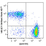
C57BL/6 mouse splenocytes were stained with CD19 (clone 6D5) Brilliant Violet 421™ (filled histogram) or rat IgG2a, κ Brilliant Violet 421™ isotype control (open histogram). -

C57BL/6 mouse frozen spleen section was fixed with 4% paraformaldehyde (PFA) for 10 minutes at room temperature and blocked with 5% FBS plus 5% rat serum for 1 hour at room temperature. Then the section was stained with 2.5 µg/ml of CD19 (clone 6D5) Brilliant Violet 421TM, 2.5 µg/ml of CD3 (clone 17A2) Alexa Fluor® 594 (red) and 2.5 µg/ml of CD11b (clone M1/70) Alexa Fluor® 488 (green) overnight at 4°C. The image was captured by 10X objective.
CD19 is a 95 kD glycoprotein also known as B4. It is a member of the Ig superfamily, expressed on all pro-B to mature B cells (during development) and follicular dendritic cells. Plasma cells do not express CD19. CD19, in association with CD21 and CD81, forms a molecular complex integral to B cell activation.
Product DetailsProduct Details
- Verified Reactivity
- Mouse
- Antibody Type
- Monoclonal
- Host Species
- Rat
- Immunogen
- Mouse CD19-expressing K562 human erythroleukemia cells
- Formulation
- Phosphate-buffered solution, pH 7.2, containing 0.09% sodium azide and BSA (origin USA).
- Preparation
- The antibody was purified by affinity chromatography and conjugated with Brilliant Violet 421™ under optimal conditions.
- Concentration
- µg sizes: 0.2 mg/mLµL sizes: lot-specific (to obtain lot-specific concentration and expiration, please enter the lot number in our Certificate of Analysis online tool.)
- Storage & Handling
- The antibody solution should be stored undiluted between 2°C and 8°C, and protected from prolonged exposure to light. Do not freeze.
- Application
-
FC - Quality tested
IHC-F - Verified - Recommended Usage
-
Each lot of this antibody is quality control tested by immunofluorescent staining with flow cytometric analysis. For immunofluorescent staining using the µg size, the suggested use of this reagent is ≤ 0.25 µg per million cells in 100 µl volume. For immunofluorescent staining using µl sizes, the suggested use of this reagent is 5 µl per million cells in 100 µl staining volume or 5 µl per 100 µl of whole blood. For immunohistochemistry on Frozen tissue using the µg size, a concentration range of 1.0 - 5.0 µg/ml is suggested. It is recommended that the reagent be titrated for optimal performance for each application.
Brilliant Violet 421™ excites at 405 nm and emits at 421 nm. The standard bandpass filter 450/50 nm is recommended for detection. Brilliant Violet 421™ is a trademark of Sirigen Group Ltd.
Learn more about Brilliant Violet™.
This product is subject to proprietary rights of Sirigen Inc. and is made and sold under license from Sirigen Inc. The purchase of this product conveys to the buyer a non-transferable right to use the purchased product for research purposes only. This product may not be resold or incorporated in any manner into another product for resale. Any use for therapeutics or diagnostics is strictly prohibited. This product is covered by U.S. Patent(s), pending patent applications and foreign equivalents. - Excitation Laser
-
Violet Laser (405 nm)
- Application Notes
-
Additional reported applications (for the relevant formats) include: immunofluorescence7.
-
Application References
(PubMed link indicates BioLegend citation) -
- Shoham T, et al. 2003. J. Immunol. 171:4062. (FC)
- Goodyear CS, et al. 2004. J. Immunol. 172:2870. (FC)
- Kamimura D, et al. 2006. J. Immunol. 177:306. (FC)
- Andoniou CE, et al. 2005. Nat. Immunol. 6:1011. (FC)
- Lawson BR, et al. 2007. J. Immunol. 178:5366. (FC)
- Phan TG, et al. 2007. Nat. Immunol. 8:992. (FC)
- Hayashida K, et al. 2008. J. Biol. Chem. 283:19895. (IF) PubMed
- Charles N, et al. 2010. Nat. Med. 16:701. (FC) PubMed
- Bankoti J, et al. 2010. Toxicol. Sci. 115:422. (FC) PubMed
- Stadnisky MD, et al. 2011. Blood. 117:5133. (FC) PubMed
- Perlot T, et al. 2012. J. Immunol. 188:1201. (FC) PubMed
- Olive V, et al. 2013. Elife. 2:822. PubMed
- Miyai T, et al. 2014. PNAS. 111:11780. PubMed
- Product Citations
-
- RRID
-
AB_10895761 (BioLegend Cat. No. 115537)
AB_2563066 (BioLegend Cat. No. 115549)
AB_11203527 (BioLegend Cat. No. 115538)
Antigen Details
- Structure
- Ig superfamily, associates with CD21 and CD81, 95 kD
- Distribution
-
Pro-B cells to mature B cells (during development), follicular dendritic cells
- Function
- Modulates B cell activation and differentiation
- Ligand/Receptor
- CD21, CD81, Leu-13
- Cell Type
- B cells, Dendritic cells
- Biology Area
- Costimulatory Molecules, Immunology
- Molecular Family
- CD Molecules
- Antigen References
-
1. Fearon DT. 1993. Curr. Opin. Immunol. 5:341.
2. Krop I, et al. 1996. Eur. J. Immunol. 26:238.
3. Krop I, et al. 1996. J. Immunol. 157:48.
4. Tedder TF, et al. 1994. Immunol. Today 15:437. - Gene ID
- 12478 View all products for this Gene ID
- UniProt
- View information about CD19 on UniProt.org
Related Pages & Pathways
Pathways


Related FAQs
- What is the F/P ratio range of our BV421™ format antibody reagents?
-
It is lot-specific. On average it ranges between 2-4.
Other Formats
View All CD19 Reagents Request Custom ConjugationCustomers Also Purchased
Compare Data Across All Formats
This data display is provided for general comparisons between formats.
Your actual data may vary due to variations in samples, target cells, instruments and their settings, staining conditions, and other factors.
If you need assistance with selecting the best format contact our expert technical support team.
-
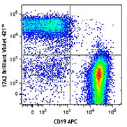
APC anti-mouse CD19

C57BL/6 mouse splenocytes stained with CD19 (clone 6D5) APC ... -
Biotin anti-mouse CD19

C57BL/6 mouse splenocytes were stained with biotinylated CD1... -
FITC anti-mouse CD19

C57BL/6 splenocytes were stained with CD19 (clone 6D5) FITC ... -
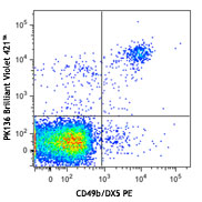
PE anti-mouse CD19

C57BL/6 mouse splenocytes were stained with CD19 (clone 6D5)... 
C57BL/6 mouse splenocytes were stained with CD19 (clone 6D5)... -
PE/Cyanine5 anti-mouse CD19

C57BL/6 splenocytes were stained with CD19 (clone 6D5) PE/Cy... -
Purified anti-mouse CD19

C57BL/6 mouse splenocytes were stained with purified CD19 (c... -
PE/Cyanine7 anti-mouse CD19

C57BL/6 splenocytes were stained with CD19 (clone 6D5) PE/Cy... -
Alexa Fluor® 488 anti-mouse CD19

C57BL/6 mouse splenocytes were stained with CD19 (clone 6D5)... -
Alexa Fluor® 647 anti-mouse CD19

C57BL/6 mouse splenocytes were stained with CD19 (clone 6D5)... 
C57 mouse frozen spleen section was fixed with 4% paraformal... -
Pacific Blue™ anti-mouse CD19

C57BL/6 mouse splenocytes were stained with CD19 (clone 6D5)... -
Alexa Fluor® 700 anti-mouse CD19

C57BL/6 mouse splenocytes were stained with CD19 (clone 6D5)... -
APC/Cyanine7 anti-mouse CD19

C57BL/6 splenocytes were stained with CD3 FITC and CD19 (clo... -
PerCP anti-mouse CD19

C57BL/6 mouse splenocytes were stained with CD19 (clone 6D5)... -
PerCP/Cyanine5.5 anti-mouse CD19

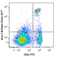
C57BL/6 mouse splenocytes were stained with CD3ε FIT... -
Alexa Fluor® 594 anti-mouse CD19

C57BL/6 mouse frozen lymph node section was fixed with 4% pa... 
C57BL/6 mouse splenocytes were stained with CD19 (clone 6D5)... -
Brilliant Violet 421™ anti-mouse CD19

C57BL/6 mouse splenocytes were stained with CD19 (clone 6D5)... 
C57BL/6 mouse frozen spleen section was fixed with 4% parafo... -
Brilliant Violet 570™ anti-mouse CD19


-
Brilliant Violet 605™ anti-mouse CD19

C57BL/6 mouse splenocytes were stained with CD19 (clone 6D5)... -
Brilliant Violet 650™ anti-mouse CD19

C57BL/6 mouse splenocytes were stained with CD19 (clone 6D5)... -
Brilliant Violet 785™ anti-mouse CD19

C57BL/6 mouse splenocytes were stained with CD19 (clone 6D5)... -
Brilliant Violet 510™ anti-mouse CD19

C57BL/6 mouse splenocytes were stained with CD19 (clone 6D5)... -
Purified anti-mouse CD19 (Maxpar® Ready)

Mouse splenocytes stained with 169Tm-anti-TCRβ (H57-597) and... -
PE/Dazzle™ 594 anti-mouse CD19

C57BL/6 mouse splenocytes were stained with CD19 (clone 6D5)... -
Brilliant Violet 711™ anti-mouse CD19

C57BL/6 mouse splenocytes were stained with CD19 (clone 6D5)... -
APC/Fire™ 750 anti-mouse CD19

C57BL/6 splenocytes were stained with CD3 PE and CD19 (clone... 
-
TotalSeq™-A0093 anti-mouse CD19
-
Brilliant Violet 750™ anti-mouse CD19

C57BL/6 mouse splenocytes were stained with CD3e PE and CD19... -
TotalSeq™-B0093 anti-mouse CD19
-
Spark Blue™ 550 anti-mouse CD19

C57BL/6 mouse splenocytes were stained with CD19 (clone 6D5)... -
Spark NIR™ 685 anti-mouse CD19

C57BL/6 mouse splenocytes were stained with CD19 (clone 6D5)... -
TotalSeq™-C0093 anti-mouse CD19
-
Ultra-LEAF™ Purified anti-mouse CD19

C57BL/6 mouse splenocytes were stained with Ultra-LEAF™ puri... -
PE/Fire™ 640 anti-mouse CD19 Antibody

): C57BL/6 mouse splenocytes were stained with anti-mouse CD... -
Spark YG™ 581 anti-mouse CD19

C57BL/6 mouse splenocytes were stained with anti-mouse CD3 P... -
APC/Fire™ 810 anti-mouse CD19

C57BL/6 mouse splenocytes were stained with anti-mouse CD3 F... -
Spark YG™ 570 anti-mouse CD19

C57BL/6 mouse frozen spleen section was fixed with 4% parafo... -
Spark Blue™ 574 anti-mouse CD19 Antibody

C57BL/6 splenocytes were stained with anti-mouse CD3e APC an... -
Spark Blue™ 515 anti-mouse CD19

C57BL/6 mouse splenocytes were stained with anti-mouse CD3ε ... -
Spark UV™ 387 anti-mouse CD19

C57BL/6 mouse splenocytes were stained with anti-mouse CD3ε ... -
Spark Red™ 718 anti-mouse CD19 (Flexi-Fluor™)


Follow Us