


- Clone
- 6D5 (See other available formats)
- Regulatory Status
- RUO
- Other Names
- B4
- Isotype
- Rat IgG2a, κ
- Ave. Rating
- Submit a Review
- Product Citations
- publications

-

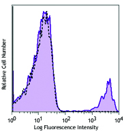
C57BL/6 mouse splenocytes were stained with biotinylated CD19 (clone 6D5) (filled histogram) or rat IgG2a, κ isotype control (open histogram), followed by Sav-PE.
| Cat # | Size | Price | Save |
|---|---|---|---|
| 115503 | 50 µg | ¥11,880 | |
| 115504 | 500 µg | ¥29,480 |
CD19 is a 95 kD glycoprotein also known as B4. It is a member of the Ig superfamily, expressed on all pro-B to mature B cells (during development) and follicular dendritic cells. Plasma cells do not express CD19. CD19, in association with CD21 and CD81, forms a molecular complex integral to B cell activation.
Product DetailsProduct Details
- Verified Reactivity
- Mouse
- Antibody Type
- Monoclonal
- Host Species
- Rat
- Immunogen
- Mouse CD19-expressing K562 human erythroleukemia cells
- Formulation
- Phosphate-buffered solution, pH 7.2, containing 0.09% sodium azide.
- Preparation
- The antibody was purified by affinity chromatography, and conjugated with biotin under optimal conditions.
- Concentration
- 0.5 mg/ml
- Storage & Handling
- The antibody solution should be stored undiluted between 2°C and 8°C. Do not freeze.
- Application
-
FC - Quality tested
- Recommended Usage
-
Each lot of this antibody is quality control tested by immunofluorescent staining with flow cytometric analysis. For flow cytometric staining, the suggested use of this reagent is ≤1.0 µg per million cells in 100 µl volume. It is recommended that the reagent be titrated for optimal performance for each application.
- Application Notes
-
Additional reported applications (for the relevant formats) include: immunofluorescence7.
-
Application References
(PubMed link indicates BioLegend citation) -
- Shoham T, et al. 2003. J. Immunol. 171:4062. (FC)
- Goodyear CS, et al. 2004. J. Immunol. 172:2870. (FC)
- Kamimura D, et al. 2006. J. Immunol. 177:306. (FC)
- Andoniou CE, et al. 2005. Nat. Immunol. 6:1011. (FC)
- Lawson BR, et al. 2007. J. Immunol. 178:5366. (FC)
- Phan TG, et al. 2007. Nat. Immunol. 8:992. (FC)
- Hayashida K, et al. 2008. J. Biol. Chem. 283:19895. (IF) PubMed
- Charles N, et al. 2010. Nat. Med. 16:701. (FC) PubMed
- Bankoti J, et al. 2010. Toxicol. Sci. 115:422. (FC) PubMed
- Stadnisky MD, et al. 2011. Blood. 117:5133. (FC) PubMed
- Perlot T, et al. 2012. J. Immunol. 188:1201. (FC) PubMed
- Olive V, et al. 2013. Elife. 2:822. PubMed
- Miyai T, et al. 2014. PNAS. 111:11780. PubMed
- Product Citations
-
- RRID
-
AB_313638 (BioLegend Cat. No. 115503)
AB_313639 (BioLegend Cat. No. 115504)
Antigen Details
- Structure
- Ig superfamily, associates with CD21 and CD81, 95 kD
- Distribution
-
Pro-B cells to mature B cells (during development), follicular dendritic cells
- Function
- Modulates B cell activation and differentiation
- Ligand/Receptor
- CD21, CD81, Leu-13
- Cell Type
- B cells, Dendritic cells
- Biology Area
- Costimulatory Molecules, Immunology
- Molecular Family
- CD Molecules
- Antigen References
-
1. Fearon DT. 1993. Curr. Opin. Immunol. 5:341.
2. Krop I, et al. 1996. Eur. J. Immunol. 26:238.
3. Krop I, et al. 1996. J. Immunol. 157:48.
4. Tedder TF, et al. 1994. Immunol. Today 15:437. - Gene ID
- 12478 View all products for this Gene ID
- UniProt
- View information about CD19 on UniProt.org
Related Pages & Pathways
Pathways


Related FAQs
- How many biotin molecules are per antibody structure?
- We don't routinely measure the number of biotins with our antibody products but the number of biotin molecules range from 3-6 molecules per antibody.
Other Formats
View All CD19 Reagents Request Custom ConjugationCustomers Also Purchased
Compare Data Across All Formats
This data display is provided for general comparisons between formats.
Your actual data may vary due to variations in samples, target cells, instruments and their settings, staining conditions, and other factors.
If you need assistance with selecting the best format contact our expert technical support team.
-
APC anti-mouse CD19

C57BL/6 mouse splenocytes stained with CD19 (clone 6D5) APC ... -
Biotin anti-mouse CD19

C57BL/6 mouse splenocytes were stained with biotinylated CD1... -
FITC anti-mouse CD19

C57BL/6 splenocytes were stained with CD19 (clone 6D5) FITC ... -
PE anti-mouse CD19

C57BL/6 mouse splenocytes were stained with CD19 (clone 6D5)... 
C57BL/6 mouse splenocytes were stained with CD19 (clone 6D5)... -
PE/Cyanine5 anti-mouse CD19

C57BL/6 splenocytes were stained with CD19 (clone 6D5) PE/Cy... -
Purified anti-mouse CD19

C57BL/6 mouse splenocytes were stained with purified CD19 (c... -
PE/Cyanine7 anti-mouse CD19

C57BL/6 splenocytes were stained with CD19 (clone 6D5) PE/Cy... -
Alexa Fluor® 488 anti-mouse CD19

C57BL/6 mouse splenocytes were stained with CD19 (clone 6D5)... -
Alexa Fluor® 647 anti-mouse CD19

C57BL/6 mouse splenocytes were stained with CD19 (clone 6D5)... 
C57 mouse frozen spleen section was fixed with 4% paraformal... -
Pacific Blue™ anti-mouse CD19

C57BL/6 mouse splenocytes were stained with CD19 (clone 6D5)... -
Alexa Fluor® 700 anti-mouse CD19

C57BL/6 mouse splenocytes were stained with CD19 (clone 6D5)... -
APC/Cyanine7 anti-mouse CD19

C57BL/6 splenocytes were stained with CD3 FITC and CD19 (clo... -
PerCP anti-mouse CD19

C57BL/6 mouse splenocytes were stained with CD19 (clone 6D5)... -
PerCP/Cyanine5.5 anti-mouse CD19

C57BL/6 mouse splenocytes were stained with CD3ε FIT... -
Alexa Fluor® 594 anti-mouse CD19

C57BL/6 mouse frozen lymph node section was fixed with 4% pa... 
C57BL/6 mouse splenocytes were stained with CD19 (clone 6D5)... -
Brilliant Violet 421™ anti-mouse CD19

C57BL/6 mouse splenocytes were stained with CD19 (clone 6D5)... 
C57BL/6 mouse frozen spleen section was fixed with 4% parafo... -
Brilliant Violet 570™ anti-mouse CD19


-
Brilliant Violet 605™ anti-mouse CD19

C57BL/6 mouse splenocytes were stained with CD19 (clone 6D5)... -
Brilliant Violet 650™ anti-mouse CD19

C57BL/6 mouse splenocytes were stained with CD19 (clone 6D5)... -
Brilliant Violet 785™ anti-mouse CD19

C57BL/6 mouse splenocytes were stained with CD19 (clone 6D5)... -
Brilliant Violet 510™ anti-mouse CD19

C57BL/6 mouse splenocytes were stained with CD19 (clone 6D5)... -
Purified anti-mouse CD19 (Maxpar® Ready)

Mouse splenocytes stained with 169Tm-anti-TCRβ (H57-597) and... -
PE/Dazzle™ 594 anti-mouse CD19

C57BL/6 mouse splenocytes were stained with CD19 (clone 6D5)... -
Brilliant Violet 711™ anti-mouse CD19

C57BL/6 mouse splenocytes were stained with CD19 (clone 6D5)... -
APC/Fire™ 750 anti-mouse CD19

C57BL/6 splenocytes were stained with CD3 PE and CD19 (clone... 
-
TotalSeq™-A0093 anti-mouse CD19
-
Brilliant Violet 750™ anti-mouse CD19

C57BL/6 mouse splenocytes were stained with CD3e PE and CD19... -
TotalSeq™-B0093 anti-mouse CD19
-
Spark Blue™ 550 anti-mouse CD19

C57BL/6 mouse splenocytes were stained with CD19 (clone 6D5)... -
Spark NIR™ 685 anti-mouse CD19

C57BL/6 mouse splenocytes were stained with CD19 (clone 6D5)... -
TotalSeq™-C0093 anti-mouse CD19
-
Ultra-LEAF™ Purified anti-mouse CD19

C57BL/6 mouse splenocytes were stained with Ultra-LEAF™ puri... -
PE/Fire™ 640 anti-mouse CD19 Antibody

): C57BL/6 mouse splenocytes were stained with anti-mouse CD... -
Spark YG™ 581 anti-mouse CD19

C57BL/6 mouse splenocytes were stained with anti-mouse CD3 P... -
APC/Fire™ 810 anti-mouse CD19

C57BL/6 mouse splenocytes were stained with anti-mouse CD3 F... -
Spark YG™ 570 anti-mouse CD19

C57BL/6 mouse frozen spleen section was fixed with 4% parafo... -
Spark Blue™ 574 anti-mouse CD19 Antibody

C57BL/6 splenocytes were stained with anti-mouse CD3e APC an... -
Spark Blue™ 515 anti-mouse CD19

C57BL/6 mouse splenocytes were stained with anti-mouse CD3ε ... -
Spark UV™ 387 anti-mouse CD19

C57BL/6 mouse splenocytes were stained with anti-mouse CD3ε ... -
Spark Red™ 718 anti-mouse CD19 (Flexi-Fluor™)


Follow Us