


- Clone
- SMI 311 (See other available formats)
- Regulatory Status
- RUO
- Other Names
- Neurofilament Marker
- Previously
-
Covance Catalog# SMI-311R
- Isotype
- Mouse IgG1/Mouse IgM
- Ave. Rating
- Submit a Review
- Product Citations
- publications

-

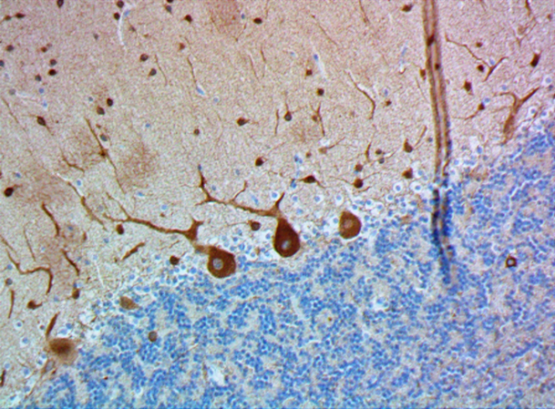
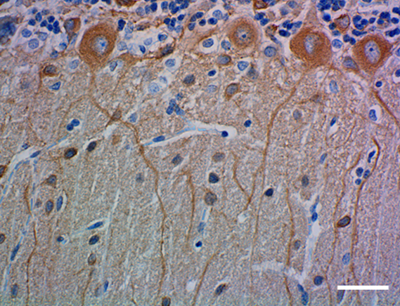
IHC staining of anti-Neurofilament Marker (pan-neuronal, cocktail) antibody (clone SMI 311) on formalin-fixed paraffin-embedded mouse brain tissue. Following antigen retrieval using Retrieve-All Antigen Unmasking System 3: Acidic, 100X (Cat. No. 927601), the tissue was incubated with a 1:5000 dilution of the primary antibody overnight at 4°C. BioLegend´s Ultra-Streptavidin (USA) HRP kit (Multi-Species, DAB, Cat. No. 929901) was used for detection followed by hematoxylin counterstaining, according to the protocol provided. The image was captured with a 40X objective. Scale bar: 50 µm -

IHC staining of anti-Neurofilament Marker (pan-neuronal, cocktail) antibody (clone SMI 311) on formalin-fixed paraffin-embedded rat brain tissue. Following antigen retrieval using Retrieve-All Antigen Unmasking System 3: Acidic, 100X (Cat. No. 927601), the tissue was incubated with a 1:5000 dilution of the primary antibody overnight at 4°C. BioLegend´s Ultra-Streptavidin (USA) HRP kit (Multi-Species, DAB, Cat. No. 929901) was used for detection followed by hematoxylin counterstaining, according to the protocol provided. The image was captured with a 40X objective. Scale bar: 50 µm -

IHC staining of anti-Neurofilament Marker (pan-neuronal, cocktail) antibody (clone SMI 311) on formalin-fixed paraffin-embedded human brain tissue. Following antigen retrieval using Retrieve-All Antigen Unmasking System 3: Acidic, 100X (Cat. No. 927601), the tissue was incubated with a 1:5000 dilution of the primary antibody overnight at 4°C. BioLegend's Ultra-Streptavidin (USA) HRP kit (Multi-Species, DAB, Cat. No. 929901) was used for detection followed by hematoxylin counterstaining, according to the protocol provided. The image was captured with a 40X objective. Scale bar: 50 µm -

IHC staining of anti-Neurofilament Marker (pan-neuronal, cocktail) antibody (clone SMI 311) on formalin-fixed paraffin-embedded mouse brain tissue. Following antigen retrieval using Retrieve-All Antigen Unmasking System 3: Acidic, 100X (Cat. No. 927601), the tissue was incubated with a 1:5000 dilution of the primary antibody overnight at 4°C, followed by incubation with Alexa Fluor® 647 Goat anti-mouse IgG (Cat. No. 405322) for one hour at room temperature. Nuclei were counterstained with DAPI, and the slide was mounted with ProLong™ Gold Antifade Mountant. The image was captured with a 40X objective. Scale Bar: 50 µm -

IHC staining of anti-Neurofilament Marker (pan-neuronal, cocktail) antibody (clone SMI 311) on formalin-fixed paraffin-embedded rat brain tissue. Following antigen retrieval using Retrieve-All Antigen Unmasking System 3: Acidic, 100X (Cat. No. 927601), the tissue was incubated with a 1:5000 dilution of the primary antibody overnight at 4°C, followed by incubation with Alexa Fluor® 647 Goat anti-mouse IgG (Cat. No. 405322) for one hour at room temperature. Nuclei were counterstained with DAPI, and the slide was mounted with ProLong™ Gold Antifade Mountant. The image was captured with a 40X objective. Scale Bar: 50 µm -

Western blot of anti-Neurofilament Marker (pan-neuronal, cocktail) antibody (clone SMI 311). Lane 1: Molecular weight marker; Lane 2: 30 µg of human brain lysate; Lane 3: 30 µg of mouse brain lysate; Lane 4: 30 µg of rat brain lysate. The blot was incubated with a 1:10,000 dilution of the primary antibody overnight at 4°C, followed by incubation with HRP labeled goat anti-mouse IgG (Cat. No. 405306). Enhanced chemiluminescence was used as the detection system.
| Cat # | Size | Price | Save |
|---|---|---|---|
| 837801 | 100 µL | ¥82,500 | |
| 837802 | 500 µL | ¥148,060 |
Neurofilaments (NF) are approximately 10 nanometer intermediate filaments found in neurons. They are a major component of the neuronal cytoskeleton and their function is primarily to provide structural support for the axon and to regulate axon diameter. There are three major NF subunits, and the names given to these subunits are based upon the apparent molecular mass of the mammalian subunits on SDS-PAGE. The light or lowest (NF-L) runs at 68-70 kD, the medium or middle (NF-M) runs at about 145-160 kD, and the heavy or highest (NF-H) runs at 200-220 kD. However, the actual molecular weight of these proteins is considerably lower due to the highly charged C-terminal regions of the molecules. The level of NF gene expression correlates with the axonal diameter, which controls how fast electrical signals travel down the axon. Mutant mice with NF abnormalities have phenotypes resembling amyotrophic lateral sclerosis. NF immunostaining is common in diagnostic neuropathology. It is useful for differentiating neurons (positive for NF) from glia (negative for NF).
Product DetailsProduct Details
- Verified Reactivity
- Human, Mouse, Rat
- Antibody Type
- Monoclonal
- Host Species
- Mouse
- Formulation
- Ascites Fluid (contains 0.01M sodium azide).
- Preparation
- Ascites
- Concentration
- The concentration is not quantified as this product is sold as undiluted crude mouse ascites fluid. The concentration might vary from lot-to-lot and an estimated concentration would be 1-3 mg/ml.
- Storage & Handling
- Store at -20°C. Upon initial thawing, apportion into working aliquots and store at -20°C. Avoid repeated freeze-thaw cycles to prevent denaturing the antibody. For long-term storage, keep the antibody at -80°C.
- Application
-
IHC-P - Quality tested
WB - Verified
IHC-F, ICC - Reported in the literature, not verified in house - Recommended Usage
-
Each lot of this antibody is quality control tested by formalin-fixed paraffin-embedded immunohistochemical staining. For immunohistochemistry, a dilution range of 1:1000 - 1:5000 is suggested. For Western blotting, the suggested use of this reagent is 1:5000 - 1:10000 per ml. It is recommended that the reagent be titrated for optimal performance for each application.
- Application Notes
-
SMI 311 has been selected to provide a specific marker for neurons in tissue sections and cultures. In contrast to individual antinonphosphoneurofilaments that identify different subset of neurons and are, therefore, especially suitable for defining anatomic and functional differences in normal and pathologic neurons, SMI 311 is a convenient marker for neurons in general and their differentiation from non-neuronal cells. SMI 311 provides an early marker of neuronal migration and differentiation in human fetal development yielding Golgi-like images without the disadvantages of lack of selectivity and poor specificity of the Golgi technique. In specifically delineating cell bodies and dendrites, SMI 311 has been used to trace the "inside-out gradient" of neuron production and differentiation. Pathologic conditions, such as undernutrition affect SMI 311-visualized soma size and dendritic arborization.
-
Application References
(PubMed link indicates BioLegend citation) -
- Tsunoda I, Kuang LQ, Libbey JE, Fujinami RS. Axonal injury heralds virus-induced demyelination. Am J Path 162:1259-1269, 2003. (IHC-P)
- Ulfig N, Nickel J, Bohl J. Monoclonal antibodies SMI 311 and SMI 312 as tools to investigate the maturation of nerve cells and axonal patterns in human fetal brain. Cell Tissue Res 291:433, 1998.
- Frola E, et al. 2013. PLoS One 2:e56311. (IHC-F)
- Ivanovic A, et al. 2012. J. Cell Biol. 3:337. (IHC-F, WB) PubMed
- Petzold A, et al. 2011. Brain 134:464. (IHC-P, WB) PubMed
- Del Valle L,et al. Cancer Biol Ther.9:286. (IHC-P, ICC, WB) PubMed
- Product Citations
-
- RRID
-
AB_2565383 (BioLegend Cat. No. 837801)
AB_2565384 (BioLegend Cat. No. 837802)
Antigen Details
- Structure
- Expected MW: medium or middle NF (NF-M) runs at 145-160 kD, and the heavy or highest NF (NF-H) runs at 200-220 kD
- Cell Type
- Mature Neurons
- Biology Area
- Cell Biology, Neuroscience, Neuroscience Cell Markers
- Molecular Family
- Intermediate Filaments
- Gene ID
- 4747 View all products for this Gene ID 4744 View all products for this Gene ID 4741 View all products for this Gene ID
- UniProt
- View information about Neurofilament Marker on UniProt.org


Related FAQs
Other Formats
View All Neurofilament Marker Reagents Request Custom Conjugation| Description | Clone | Applications |
|---|---|---|
| Anti-Neurofilament Marker (pan-neuronal, cocktail) | SMI 311 | IHC-P,WB,IHC-F,ICC |
Customers Also Purchased

Compare Data Across All Formats
This data display is provided for general comparisons between formats.
Your actual data may vary due to variations in samples, target cells, instruments and their settings, staining conditions, and other factors.
If you need assistance with selecting the best format contact our expert technical support team.
-
Anti-Neurofilament Marker (pan-neuronal, cocktail)

IHC staining of anti-Neurofilament Marker (pan-neuronal, coc... 
IHC staining of anti-Neurofilament Marker (pan-neuronal, coc... 
IHC staining of anti-Neurofilament Marker (pan-neuronal, coc... 
IHC staining of anti-Neurofilament Marker (pan-neuronal, coc... 
IHC staining of anti-Neurofilament Marker (pan-neuronal, coc... 
Western blot of anti-Neurofilament Marker (pan-neuronal, coc...


Follow Us