


- Clone
- D8B7 (See other available formats)
- Regulatory Status
- RUO
- Other Names
- Spectrin alpha chain, brain, alpha-II spectrin, fodrin alpha chain, spectrin, non-erythroid alpha chain, EL2, SPTA1
- Previously
-
Covance Catalog# SIG-39702
Signet Catalog# 9702-02
Signet Catalog# 9702-05
Signet Catalog# 9702-10
- Isotype
- Mouse IgG2b, κ
- Ave. Rating
- Submit a Review
- Product Citations
- publications

-

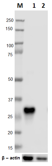
Western blot of anti-Alpha-II Spectrin antibody (clone D8B7). Lane 1: Molecular weight marker; Lane 2: 20 µg of human brain lysate; Lane 3: 20 µg of mouse brain lysate; Lane 4: 20 µg of rat brain lysate; Lane 5: 20 µg of 3T3 cell lysate; Lane 6: 20 µg of HeLa cell lysate. The blot was incubated with the primary antibody at 1:2000 dilution overnight at 4°C, followed by incubation with HRP labeled goat anti-mouse IgG (Cat. No. 405306). Enhanced chemiluminescence was used as the detection system. -

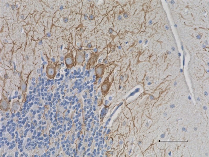
IHC staining of anti-Alpha-II Spectrin antibody (clone D8B7) on formalin-fixed paraffin-embedded mouse brain tissue. Following antigen retrieval using Sodium Citrate H.I.E.R., the tissue was incubated with the primary antibody at 1:1000 dilution overnight at 4°C. BioLegend´s Ultra-Streptavidin (USA) HRP kit (Multi-Species, DAB, Cat. No. 929901) was used for detection followed by hematoxylin counterstaining, according to the protocol provided. The image was captured with a 40X objective. Scale bar: 50 µm -

IHC staining of anti-Alpha-II Spectrin antibody (clone D8B7) on formalin-fixed paraffin-embedded rat brain tissue. Following antigen retrieval using Sodium Citrate H.I.E.R., the tissue was incubated with the primary antibody at 1:1000 dilution overnight at 4°C. BioLegend´s Ultra-Streptavidin (USA) HRP kit (Multi-Species, DAB, Cat. No. 929901) was used for detection followed by hematoxylin counterstaining, according to the protocol provided. The image was captured with a 40X objective. Scale bar: 50 µm -

ICC staining of anti-Alpha-II Spectrin antibody (clone D8B7) on HeLa cells. The cells were fixed with 4% PFA, permeabilized with a buffer containing 0.1% Triton X-100 and 0.25% BSA, and blocked with 2% normal goat serum and 0.02% BSA. The cells were then incubated with the primary antibody at 1:1000 dilution overnight at 4°C, followed by incubation with 2.5 µg/ml of Alexa Fluor® 594 goat anti-Mouse IgG for one hour at room temperature. Nuclei were counterstained with DAPI, and the slides were mounted with ProLongTM Gold Antifade Mountant. The image was captured with a 40X objective. Scale bar: 50 µm -

ICC staining of anti-Alpha-II Spectrin antibody (clone D8B7) on 3T3 cells. The cells were fixed with 4% PFA, permeabilized with a buffer containing 0.1% Triton X-100 and 0.25% BSA, and blocked with 2% normal goat serum and 0.02% BSA. The cells were then incubated with the primary antibody at 1:1000 dilution overnight at 4°C, followed by incubation with 2.5 µg/ml of Alexa Fluor® 594 goat anti-Mouse IgG for one hour at room temperature. Nuclei were counterstained with DAPI, and the slides were mounted with ProLongTM Gold Antifade Mountant. The image was captured with a 40X objective. Scale bar: 50 µm
The spectrins are a family of widely distributed cytoskeletal proteins which are involved in actin crosslinking. Spectrin α-chain heterodimerizes with tissue-specific β-chains. It is specifically expressed in nonerythrocytic cells. The protein has been implicated in other cellular functions including DNA repair and cell cycle regulation. Mutations in the gene are the cause of early infantile epileptic encephalopathy. Fodrin, a spectrin that seems to be involved in secretion, interacts with calmodulin in a calcium-dependent manner and is thus a candidate for the calcium-dependent movement of the cytoskeleton at the membrane.
Product DetailsProduct Details
- Verified Reactivity
- Human, Mouse, Rat, Drosophila
- Antibody Type
- Monoclonal
- Host Species
- Mouse
- Preparation
- Ascites
- Storage & Handling
- Store at -20°C or below. Upon initial thawing, apportion into working aliquots and store at -20°C or below. Avoid repeated freeze-thaw cycles to prevent denaturing the antibody. Do not store in frost-free freezers.
- Application
-
WB - Quality tested
IHC-P, ICC - Verified - Recommended Usage
-
Each lot of this antibody is quality control tested by Western blotting. For Western blotting, the suggested use of this reagent is 1:2000 - 1:4000 dilution. For immunohistochemistry on formalin-fixed paraffin-embedded tissue, a dilution of 1:1000 is suggested. For immunocytochemistry, a dilution of 1:1000 is recommended. It is recommended that the reagent be titrated for optimal performance for each application.
- Application Notes
-
Clone D8B7 is a specific marker for human, rat and mouse alpha II spectrin or fodrin. In cerebellar cell cultures, it labels axons and periphery of cell bodies consistent with localization of fodrin (reviewed in Bennett, 1993; Goodman, 1995 & De Matteis, 2000)4, 6, 8. It does not cross-react with alpha I (erythroid) spectrin SH3 domain.
This antibody was developed against a recombinant alpha II spectrin (non-erythroid spectrin or fodrin) SH3 domain. It was subsequently purified via ammonium sulfate precipitation.
Reactivity to Drosophila was only verified with the purified format. -
Application References
(PubMed link indicates BioLegend citation) -
- Konieczny P, et al. 2008. J Cell Biol. 181:667-81
- Xu J, et al. 2001. Brain Res. 898:171-7 (WB, IF)
- Xu J, et al. 2000. J Cell Science. 113:3805-14
- De Matteis MA and Morrow JS. 2000. J. Cell Science. 113:2331-43
- Swanson JA and Watts C. 1995. Trends Cell Biol. 5:424-427
- Goodman SR, et al. 1995. Brain Res Bull. 36:593-606
- Lamaze C and Schimid SL. 1995. Curr Opin Cell Biol. 7:573-580
- Bennett V and Gilligan DM. 1993. Annu Rev Cell Biol. 9:27-66
- Huang C, et al. 2017. J Neurosci. 37:11323-11334 PubMed
- Product Citations
-
- RRID
-
AB_2564660 (BioLegend Cat. No. 803201)
Antigen Details
- Structure
- Alpha-II Spectrin is a 2472 amino acid protein with an expected molecular mass of 285 kD. A cleavage product of ~ 150 kD is observed in tissue lysates (1) (2).
- Biology Area
- Cell Biology, Cell Motility/Cytoskeleton/Structure, Neuroscience, Neuroscience Cell Markers
- Molecular Family
- Presynaptic proteins, Scaffold Proteins
- Antigen References
-
- Pike BR, et al. 2001. J Neurochem. 78(6):1297-306.
- Weiss ES, et al. 2009. Ann Thorac Surg. 88(2):543
- Gene ID
- 6709 View all products for this Gene ID
- UniProt
- View information about alpha-II Spectrin on UniProt.org


Related FAQs
Other Formats
View All Alpha-II Spectrin Reagents Request Custom Conjugation| Description | Clone | Applications |
|---|---|---|
| Anti-Alpha-II Spectrin | D8B7 | WB,IHC-P,ICC |
| Purified anti-Alpha-II Spectrin | D8B7 | IHC-P,ICC,WB |
| HRP anti-Alpha-II Spectrin | D8B7 | WB,IHC-P |
| Biotin anti-Alpha-II Spectrin | D8B7 | WB,IHC-P |
| Alexa Fluor® 647 anti-Alpha-II Spectrin | D8B7 | IHC-P |
Customers Also Purchased
Compare Data Across All Formats
This data display is provided for general comparisons between formats.
Your actual data may vary due to variations in samples, target cells, instruments and their settings, staining conditions, and other factors.
If you need assistance with selecting the best format contact our expert technical support team.
-
Anti-Alpha-II Spectrin

Western blot of anti-Alpha-II Spectrin antibody (clone D8B7)... 
IHC staining of anti-Alpha-II Spectrin antibody (clone D8B7)... 
IHC staining of anti-Alpha-II Spectrin antibody (clone D8B7)... 
ICC staining of anti-Alpha-II Spectrin antibody (clone D8B7)... 
ICC staining of anti-Alpha-II Spectrin antibody (clone D8B7)... -
Purified anti-Alpha-II Spectrin

IHC staining of purified anti-Alpha-II Spectrin antibody (cl... 
IHC staining of purified anti-Alpha-II Spectrin antibody (cl... 
ICC staining of purified anti-Alpha-II Spectrin antibody (cl... 
ICC staining of purified anti-Alpha-II Spectrin antibody (cl... 
Western blot of purified anti-Alpha-II Spectrin antibody (cl... 
Western blot of purified anti-Alpha-II Spectrin antibody (cl... -
HRP anti-Alpha-II Spectrin

Western blot of HRP anti-Alpha-II-Spectrin antibody (clone D... 
IHC staining of HRP anti-Alpha-II-Spectrin antibody (clone D... 
IHC staining of HRP anti-Alpha-II-Spectrin antibody (clone D... 
IHC staining of HRP anti-Alpha-II-Spectrin antibody (clone D... -
Biotin anti-Alpha-II Spectrin

Western blot of Biotin anti-Alpha-II-Spectrin antibody (clon... 
IHC staining of Biotin anti-Alpha-II-Spectrin antibody (clon... 
IHC staining of Biotin anti-Alpha-II-Spectrin antibody (clon... 
IHC staining of Biotin anti-Alpha-II-Spectrin antibody (clon... -
Alexa Fluor® 647 anti-Alpha-II Spectrin

IHC staining of Alexa Fluor® 647 anti-Alpha-II Spectrin anti... 
IHC staining of Alexa Fluor® 647 anti-Alpha-II Spectrin anti... 
IHC staining of Alexa Fluor® 647 anti-Alpha-II Spectrin anti... 
IHC staining of Alexa Fluor® 647 anti-Alpha-II Spectrin anti...


Follow Us