 Login / Register


- Clone
- 12G5 (See other available formats)
- Regulatory Status
- RUO
- Workshop
- VII 70204
- Other Names
- CXCR4, Fusin
- Isotype
- Mouse IgG2a, κ
- Ave. Rating
- Submit a Review
- Product Citations
- publications

-

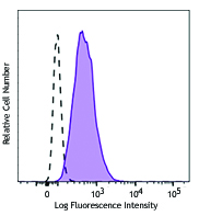
Human peripheral blood lymphocytes were stained with CD3 FITC and CD184 (CXCR4) (clone 12G5) Brilliant Violet 510™ (left) or mouse IgG2a, κ isotype control Brilliant Violet 510™ (right).
| Cat # | Size | Price | Quantity Check Availability | Save | ||
|---|---|---|---|---|---|---|
| 306535 | 25 tests | 168€ | ||||
| 306536 | 100 tests | 352€ | ||||
CD184, also known as fusin or CXCR4, is a 45 kD seven transmembrane G-protein-linked CXC chemokine receptor. CD184 is widely expressed on blood and tissue cells, including B and T cells, monocytes, macrophages, dendritic cells, granulocytes, megakaryocytes/platelets, lymphoid, myeloid precursor cells, endothelial cells, epithelial cells, astrocytes, and neurons, among other tissue cells. CD184 is the receptor for CXC chemokine SDF-1, mediates blood cell migration, and is involved in B lymphopoiesis and myelopoiesis, cardiogenesis, blood vessel formation, and cerebellar development. CXCR4 is also a coreceptor of X4 HIV-1 and an alternative receptor for some isolates of HIV-2.
Product DetailsProduct Details
- Verified Reactivity
- Human, Cynomolgus, Rhesus
- Reported Reactivity
- African Green, Baboon, Chimpanzee, Sooty Mangabey
- Antibody Type
- Monoclonal
- Host Species
- Mouse
- Immunogen
- CP-MAC-infected Sup-T1 cells
- Formulation
- Phosphate-buffered solution, pH 7.2, containing 0.09% sodium azide and BSA (origin USA).
- Preparation
- The antibody was purified by affinity chromatography and conjugated with Brilliant Violet 510™ under optimal conditions.
- Concentration
- Lot-specific (to obtain lot-specific concentration and expiration, please enter the lot number in our Certificate of Analysis online tool.)
- Storage & Handling
- The antibody solution should be stored undiluted between 2°C and 8°C, and protected from prolonged exposure to light. Do not freeze.
- Application
-
FC - Quality tested
- Recommended Usage
-
Each lot of this antibody is quality control tested by immunofluorescent staining with flow cytometric analysis. For flow cytometric staining, the suggested use of this reagent is 5 µl per million cells in 100 µl staining volume or 5 µl per 100 µl of whole blood.
Brilliant Violet 510™ excites at 405 nm and emits at 510 nm. The bandpass filter 510/50 nm is recommended for detection, although filter optimization may be required depending on other fluorophores used. Be sure to verify that your cytometer configuration and software setup are appropriate for detecting this channel. Refer to your instrument manual or manufacturer for support. Brilliant Violet 510™ is a trademark of Sirigen Group Ltd.
Learn more about Brilliant Violet™.
This product is subject to proprietary rights of Sirigen Inc. and is made and sold under license from Sirigen Inc. The purchase of this product conveys to the buyer a non-transferable right to use the purchased product for research purposes only. This product may not be resold or incorporated in any manner into another product for resale. Any use for therapeutics or diagnostics is strictly prohibited. This product is covered by U.S. Patent(s), pending patent applications and foreign equivalents. - Excitation Laser
-
Violet Laser (405 nm)
- Application Notes
-
Additional reported applications (for the relevant formats) include: immunohistochemical staining of paraffin-embedded tissue sections11, immunocytochemistry3, immunofluorescence microscopy2,6, and blocking of CD4-independent infection by HIV-2 and CD4-dependent infection by some T cell-tropic isolates of HIV-14,5. Clone 12G5 may not be suitable for Western blotting.10 The Ultra-LEAF™ purified antibody (Endotoxin <0.01 EU/µg, Azide-Free, 0.2 µm filtered) is recommended for functional assays (Cat. Nos. 306539 & 306540).
- Application References
-
- McKnight A, et al. 1997. J. Virol. 71:1692.
- Endres MJ, et al. 1996. Cell 87:745. (Immunogen, IF)
- Volin MV, et al. 1998. Biochem. Biophys. Res. Commun. 242:46. (ICC)
- Berndt C, et al. 1998. P. Natl. Acad. Sci. USA 95:12556. (Block)
- Ullrich CK, et al. 2000. Blood 96:1438. (Block)
- Murga M, et al. 2005. Blood 105:1992. (IF)
- Thompson BD. 2007. J. Biol. Chem. 282:9547. (FC) PubMed
- Isnardi I, et al. 2010. Blood 115:5026. PubMed
- Yoshino N, et al. 2000. Exp. Anim. (Tokyo) 49:97. (FC)
- Fischer T, et al. 2008. PLoS One 3:e4069.
- Schmid BC, et al. 2004. Breast Cancer Res. Treat. 84:247. (IHC)
- RRID
-
AB_2810460 (BioLegend Cat. No. 306535)
AB_2810461 (BioLegend Cat. No. 306536)
Antigen Details
- Structure
- Rhodopsin family, G-protein linked seven transmembrane glycoprotein, 45 kD
- Distribution
-
T cells and B cells, dendritic cells, monocytes, granulocytes, hematopoietic progenitors, endothelial cells
- Function
- B lymphopoiesis and myelopoiesis, cardiogenesis, blood vessel formation, cerebellar development
- Ligand/Receptor
- SDF-1 receptor, coreceptor for X4 HIV-1
- Cell Type
- B cells, Dendritic cells, Endothelial cells, Granulocytes, Hematopoietic stem and progenitors, Mesenchymal Stem Cells, Monocytes, Neural Stem Cells, T cells, Tregs
- Biology Area
- Cell Biology, Immunology, Innate Immunity, Neuroinflammation, Neuroscience, Neuroscience Cell Markers, Stem Cells
- Molecular Family
- CD Molecules, Cytokine/Chemokine Receptors, GPCR
- Antigen References
-
1. Berger E, et al. 1999. Annu. Rev. Immunol. 17:657.
2. Loetscher P, et al. 2000. Adv. Immunol. 74:127.
3. Murphy P, et al. 2000. Pharmacol. Rev. 52:145. - Gene ID
- 7852 View all products for this Gene ID
- UniProt
- View information about CD184 on UniProt.org
Related Pages & Pathways
Pathways


Related FAQs
- Does staining at room temperature or even at 37°C help for checking chemokine receptors expression?
-
Due to continuous recycling of many chemokine receptors, it may be worthwhile to consider staining at room temperature or at 37°C if the staining at lower temperature (which can potentially reduce receptor turnover) is not optimal.
Other Formats
View All CD184 Reagents Request Custom ConjugationCustomers Also Purchased
Compare Data Across All Formats
This data display is provided for general comparisons between formats.
Your actual data may vary due to variations in samples, target cells, instruments and their settings, staining conditions, and other factors.
If you need assistance with selecting the best format contact our expert technical support team.
-
APC anti-human CD184 (CXCR4)

Human peripheral blood lymphocytes stained with 12G5 APC -
Biotin anti-human CD184 (CXCR4)

Human peripheral blood lymphocytes stained with biotinylated... -
PE anti-human CD184 (CXCR4)

Human peripheral blood lymphocytes stained with 12G5 PE -
PE/Cyanine5 anti-human CD184 (CXCR4)

Human peripheral blood lymphocytes stained with 12G5 PE/Cyan... -
Purified anti-human CD184 (CXCR4)

Human peripheral blood lymphocytes stained with purified 12G... 
MCF7 breast cancer cell line was stained with anti-human CD1... -
PerCP/Cyanine5.5 anti-human CD184 (CXCR4)

Human peripheral blood lymphocytes stained with 12G5 PerCP/C... -
PE/Cyanine7 anti-human CD184 (CXCR4)

Human peripheral blood lymphocytes stained with 12G5 PE/Cyan... -
Brilliant Violet 421™ anti-human CD184 (CXCR4)

Human peripheral blood lymphocytes were stained with CD184 (... -
Brilliant Violet 605™ anti-human CD184 (CXCR4)

Human peripheral blood lymphocytes were stained with CD184 (... -
Purified anti-human CD184 (CXCR4) (Maxpar® Ready)

Human PBMCs stained with 175Lu-anti-CD184 (CXCR4) (12G5). Da... -
PE/Dazzle™ 594 anti-human CD184 (CXCR4)

Human peripheral blood lymphocytes were stained with CD184 (... -
APC/Cyanine7 anti-human CD184 (CXCR4)

Human peripheral blood lymphocytes were stained with CD184 (... -
Brilliant Violet 785™ anti-human CD184 (CXCR4)

Human peripheral blood lymphocytes were stained with Brillia... -
TotalSeq™-A0366 anti-human CD184 (CXCR4)
-
TotalSeq™-C0366 anti-human CD184 (CXCR4)
-
Brilliant Violet 510™ anti-human CD184 (CXCR4)

Human peripheral blood lymphocytes were stained with CD3 FIT... -
Ultra-LEAF™ Purified anti-human CD184 (CXCR4)
Human peripheral blood lymphocytes stained with LEAF™ purifi... 
MCF7 breast cancer cell line was stained with anti-human CD1... -
APC/Fire™ 750 anti-human CD184 (CXCR4)

Human peripheral blood lymphocytes were stained with CD3 FIT... -
TotalSeq™-B0366 anti-human CD184 (CXCR4)
-
TotalSeq™-D0366 anti-human CD184 (CXCR4)


Follow Us