 Login / Register


- Regulatory Status
- RUO
- Other Names
- Programmed Cell Death Protein 1, PD1, PDCD1, CD2791, PD1, PDCD1, CD279PD-1, PD 1, Programmed Cell Death Protein
- Ave. Rating
- Submit a Review
- Product Citations
- publications

-

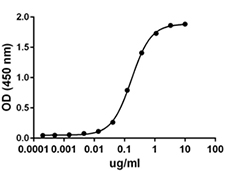
When human B7-H1 (PD-L1, CD274)-Fc chimera is immobilized at 1 μg/mL, biotinylated recombinant human PD-1 (CD279)-Fc chimera binds in a dose-dependent manner. The EC50 range for this effect is 25 - 100 ng/mL. HRP Avidin (Cat. No. 405103) was used to detect the binding. -

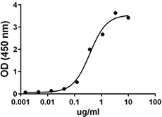
Stability Testing for Recombinant Human PD-1 (CD279)-Fc Chimera. Recombinant human PD-1 (CD279)-Fc chimera was aliquoted in PBS at 0.2 mg/mL. One aliquot was frozen and thawed four times (4x Freeze/Thaw), and compared to a control kept at 4°C (Control). The samples were tested in a binding assay with human B7-H1 (PD-L1, CD274)-Fc Chimera.
| Cat # | Size | Price | Quantity Check Availability | Save | ||
|---|---|---|---|---|---|---|
Programmed death-1 (PD-1) is a type I transmembrane protein initially isolated from apoptosis-induced cells by subtractive hybridization. The amino acid sequence of mouse PD-1 shares 59.4% identity to the human counterpart, and a putative tyrosine kinase-association motif is well conserved. Its extracellular region consists of a single Ig-like variable (IgV) domain. PD-1 is an inhibitory molecule expressed by activated B and T cells and has been implicated in immune tolerance. PD-L1 and PD-L2 are the ligands for PD-1 and their binding leads to the inhibition of T cell receptor-mediated lymphocyte proliferation and cytokine secretion. The PD-1/PD-L1 interaction suppresses immune responses against autoantigens and tumors, and plays an important role in the maintenance of peripheral immune tolerance. Interaction between PD-1 and PD-L1 promote tolerance by blocking TCR-mediated signaling. PD-L1 expression by tumor cells allow tumor progression by suppressing antitumor T-cell responses. Antibodies against PD-1 or PD-L1 lead to increased antitumor immunity.
Product DetailsProduct Details
- Source
- Human PD-1, amino acid Leu25-Gln167 (Accession # Q15116.3), with a C-terminal hIgG1 Fc tag, was expressed in 293E cells.
- Molecular Mass
- The unlabeled 382 amino acid recombinant protein has a predicted molecular mass of approximately 42.8 kD. The DTT-reduced protein migrates at approximately 60 - 70 kD and and non-reduced protein migrates at approximately 100 - 120 kD by SDS-PAGE. The predicted N-terminal amino acid is Leu.
- Purity
- > 95%, as determined by Coomassie stained SDS-PAGE
- Formulation
- 0.22 µm filtered protein solution is in PBS, 5% glycerol.
- Endotoxin Level
- Less than 0.1 EU per µg protein as determined by the LAL method.
- Concentration
- 10 and 25 µg sizes are bottled at 200 µg/mL. 100 µg size and larger sizes are lot-specific and bottled at the concentration indicated on the vial. To obtain lot-specific concentration and expiration, please enter the lot number in our Certificate of Analysis online tool.
- Storage & Handling
- Unopened vial can be stored between 2°C and 8°C for up to 2 weeks at -20°C for up to six months, or at -70°C or colder until the expiration date. For maximum results, quick spin vial prior to opening. The protein can be aliquoted and stored at -20°C or colder. Stock solutions can also be prepared at 50 - 100 µg/mL in appropriate sterile buffer, carrier protein such as 0.2 - 1% BSA or HSA can be added when preparing the stock solution. Aliquots can be stored between 2°C and 8°C for up to one week and stored at -20°C or colder for up to 3 months. Avoid repeated freeze/thaw cycles.
- Activity
- When human B7-H1 (PD-L1, CD274)-Fc chimera is immobilized at 1 µg/mL, biotinylated recombinant human PD-1 (CD279)-Fc chimera binds in a dose-dependent manner. The EC50 range for this effect is 25 - 100 ng/mL. HRP Avidin (Cat. No. 405103) was used to detect the binding.
- Application
-
Bioassay
- Application Notes
-
BioLegend carrier-free recombinant proteins provided in liquid format are shipped on blue-ice. Our comparison testing data indicates that when handled and stored as recommended, the liquid format has equal or better stability and shelf-life compared to commercially available lyophilized proteins after reconstitution. Our liquid proteins are validated in-house to maintain activity after shipping on blue ice and are backed by our 100% satisfaction guarantee. If you have any concerns, contact us at tech@biolegend.com.
Antigen Details
- Structure
- Covalently linked homodimer, biotinylated via amines
- Distribution
-
Antigen-stimulated T and B cells, regulatory T cells, follicular T and B cells, dendritic cells, and monocytes
- Function
- Inhibitory receptor on antigen activated T-cells that plays a critical role in induction and maintenance of immune tolerance to self
- Interaction
- APC, monocytes, dendritic cells, stromal cell, cancer cells
- Ligand/Receptor
- B7-H1 (PD-L1, CD274) and B7-DC (PD-L2, CD273)
- Bioactivity
- Measured by its ability to bind recombinant human B7-H1 (PD-L1, CD274)-Fc Chimera
- Cell Type
- Antigen-presenting cells, B cells, Dendritic cells, Monocytes, T cells
- Biology Area
- Adaptive Immunity, Immuno-Oncology, Immunology
- Molecular Family
- Immune Checkpoint Receptors, Soluble Receptors
- Antigen References
-
1. Ishida Y, et al. 1992. EMBO J. 11:3887-95.
2. Fife BT, et al. 2009. Nat Immunol. 10:1185-92.
3. Freeman GJ, et al. 2000. J Exp Med. 192:1027-34.
4. Latchman Y, et al. 2001. Nat Immunol. 2:261-8.
5. Iwai Y, et al. 2002. Proc Natl Acad Sci U S A. 99:12293-7.
6. Dai S, et al. 2014. Cell Immunol. 290:72-9.
7. Tan S, et al. 2017. Nature Commun. 8:14369. - Gene ID
- 5133 View all products for this Gene ID
- Specificity (DOES NOT SHOW ON TDS):
- PD-1
- Specificity Alt (DOES NOT SHOW ON TDS):
- PD-1
- App Abbreviation (DOES NOT SHOW ON TDS):
- BA
- UniProt
- View information about PD-1 on UniProt.org


Follow Us