 Login / Register


- Clone
- 143F (See other available formats)
- Regulatory Status
- RUO
- Other Names
- X-box binding protein 1
- Isotype
- Mouse IgG2a, κ
- Ave. Rating
- Submit a Review
- Product Citations
- publications

-

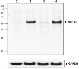
Total cell lysates (15 µg protein) from HepG2 cells treated without (-) or with (+) 300 nM thapsigargan for the indicated timepoints were resolved by 4-12% Bis-Tris gel electrophoresis, transferred to a PVDF membrane, and probed with 0.5 µg/mL (1:5000 dilution) of purified anti-Xbp-1s antibodies (clones 9D11A43 and 143F) overnight at 4°C. Proteins were visualized by chemiluminescence detection using HRP goat anti-rat IgG antibody (Cat. No. 405405) at a 1:3000 dilution. Equal protein loading was confirmed using Direct-Blot™ HRP anti-GAPDH antibody (Cat. No. 607904) used at a 1:25000 dilution (lower). Lane M: Molecular weight ladder. -

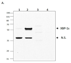
Extracts of untreated HepG2 cells (lane 1) and extracts of HepG2 cells treated with 300 nM thapsigargin (TG) for 16 hours (lane 2) were resolved by electrophoresis, transferred to nitrocellulose, and probed with purified monoclonal anti-Xbp-1s antibody (clone 143F). Proteins were visualized using a goat anti-mouse-IgG secondary conjugated to HRP and chemiluminescence detection. -

Chromatin Immunoprecipitations (ChIP) were performed using fixed and sonicated chromatin samples from 293T cells treated with tunicamycin (2.0 µg/mL, 8 hr). ChIP was performed with 10.0 µg of chromatin and 2.0 µg of purified anti- XBP-1s antibody (clone 143F) or equal amount of Go-ChIP-Grade™ purified mouse IgG2a, κ isotype ctrl antibody (Cat. No. 401505). The enriched DNA was purified and quantified by real-time qPCR using SYBR Green and primers for the human DNAJB9 exon 1 region and the human α-Satellite repeats. The amount of immunoprecipitated DNA in each sample is represented as percentage of the total amount of input chromatin, which is equivalent to 100%
| Cat # | Size | Price | Quantity Check Availability | Save | ||
|---|---|---|---|---|---|---|
| 647501 | 25 µg | 100€ | ||||
| 647502 | 100 µg | 235€ | ||||
XBP-1 is a transcription factor containing a bZIP domain. It was first identified because of its ability to bind X-box, a conserved transcriptional element in the promoter of human HLA DR gene. XBP-1 has multiple functions. It controls MHC class II gene regulation and is also essential for differentiation of plasma cells. XBP-1 upregulates as part of the ER stress response, also known as the unfolded protein response. Unspliced XBP-1 is 261 amino acids and migrates on the SDS-PAGE around 33 kD; spliced XBP-1, recognized by clone 143F, is 371 amino acids and migrates around 55 kD.
Product DetailsProduct Details
- Verified Reactivity
- Human
- Antibody Type
- Monoclonal
- Host Species
- Mouse
- Immunogen
- XBP-1s recombinant protein
- Formulation
- Phosphate-buffered solution, pH 7.2, containing 0.09% sodium azide.
- Preparation
- The antibody was purified by affinity chromatography.
- Concentration
- 0.5 mg/mL
- Storage & Handling
- The antibody solution should be stored undiluted between 2°C and 8°C.
- Application
-
WB - Quality tested
ChIP - Verified
ICC, IHC-P - Reported in the literature, not verified in house - Recommended Usage
-
Each lot of this antibody is quality control tested by Western blotting. For Western blotting, the suggested use of this reagent is 1.0 to 2.0 µg per mL. For ChIP application, use 2 µg of antibody per 10 µg of chromatin per IP. It is recommended that the reagent be titrated for optimal performance for each application.
- Application Notes
-
Additional reported applications (for the relevant formats) include: immunofluorescence14 and immunohistochemical staining of PFA-fixed paraffin-embedded sections1,14.
- Additional Product Notes
-
Using this antibody at a ratio higher than 1:5 (µg antibody per µg of chromatin) will increase background.
-
Application References
(PubMed link indicates BioLegend citation) -
- Tsang KY, et al. 2007. PLOS Biol. 5:568. (IHC) PubMed
- Farhan H, et al. 2008. EMBO J. 27:2043 (WB) PubMed
- Farhan H, et al. 2010. J. Cell Biol. 189:997. PubMed
- Feng-Jin G, et al. 2010. Cell Signal. [Epub ahead of print]
- Wang L, et al. 2011. Hum Mol Genet. PubMed
- Li J, et al. 2011. J. Biol Chem. 286:4912. PubMed
- Wang L, et al. 2011. Hum Mol Genet. 20:1008. PubMed
- Liu Y, et al. 2011. J Biol Chem. 286:13161. PubMed
- Shirley CM, et al. 2011 Blood. 117:6297. PubMed
- Vidal RL., et al. 2012. Hum Mol Genet. PubMed
- Byrd AE, et al. 2012. J Cell Biol. 196:689. PubMed
- Dickie LJ, et al. 2012. Ann rheum Dis. 71:2035. PubMed
- Bonetti P, et al. 2013. Blood. 122:2233. PubMed
- Maestre L, et al. 2009. Haematologica. 94:419. (IF, IHC)
- Rodriguez M, et al. 2014. J Biol Chem. 289:22942. PubMed
- Product Citations
-
- RRID
-
AB_2241744 (BioLegend Cat. No. 647501)
AB_2241743 (BioLegend Cat. No. 647502)
Antigen Details
- Distribution
-
Ubiquitously expressed.
- Function
- Upregulated by ER stress, IL-6, and IL-4. Downregulation correlates with tumor progression in prostate cancer. Downregulated by PAX5 transcription factor.
- Biology Area
- Cell Biology
- Antigen References
-
1. Liou HC, et al. 1990. Science 247:1581.
2. Ponath PD, et al. 1993. J. Biol. Chem.268:17074.
3. Takahashi S, et al. 2002. Prostate 50:154. - Gene ID
- 7494 View all products for this Gene ID
- UniProt
- View information about XBP-1s on UniProt.org

Other Formats
View All XBP-1s Reagents Request Custom Conjugation| Description | Clone | Applications |
|---|---|---|
| Purified anti-XBP-1s | 143F | WB,ChIP,ICC,IHC-P |
| PE anti-XBP-1s | 143F | ICFC |
| Alexa Fluor® 647 anti-XBP-1s | 143F | ICFC |
Customers Also Purchased
Compare Data Across All Formats
This data display is provided for general comparisons between formats.
Your actual data may vary due to variations in samples, target cells, instruments and their settings, staining conditions, and other factors.
If you need assistance with selecting the best format contact our expert technical support team.
-
Purified anti-XBP-1s

Extracts of untreated HepG2 cells (lane 1) and extracts of H... 
Chromatin Immunoprecipitations (ChIP) were performed using f... 
Total cell lysates (15 µg protein) from HepG2 cells treated ... -
PE anti-XBP-1s

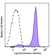
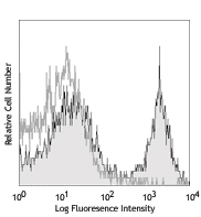
Human hepatocellular carcinoma cell line, HEPG2, was incubat... 
-
Alexa Fluor® 647 anti-XBP-1s

Human hepatocellular carcinoma cell line, HEPG2, was incubat... 


Follow Us