 Login / Register


- Clone
- IG191E/A8 (See other available formats)
- Regulatory Status
- RUO
- Other Names
- B-cell lymphoma 6, BCL-5, LAZ3, BCL6A, ZNF51, ZBTB27
- Isotype
- Mouse IgG1
- Ave. Rating
- Submit a Review
- Product Citations
- publications

-

Total lysates (15 µg protein) from Raji (lane1), HeLa (lane 2) and A20 (lane 3) cells were resolved by electrophoresis (4-20% Tris-Glycine gel), transferred to nitrocellulose, and probed with 1:500 diluted (1 µg/mL) Purified anti-Bcl6 Antibody, clone IG191E/A8 (upper). Proteins were visualized by chemiluminescence detection using a 1:3000 diluted anti-Mouse-IgG secondary antibody conjugated to HRP for the anti-Bcl6 Antibody or 1:5000 diluted Direct-Blot™ HRP anti-β-Actin Antibody, clone 2F1-1(lower). Lane M: Molecular weight ladder. -

Total cell lysates (15 µg protein) from Raji (lane 1) and HeLa cells (lane 2) were resolved by electrophoresis (4-20% Tris-Glycine gel), transferred to a nitrocellulose membrane, and probed with a 1:500 dilution (1 µg/mL) of purified anti-mouse/human Bcl-6 antibody (clone IG191E/A8). Proteins were visualized using chemiluminescence detection after incubation with a 1:3000 dilution of HRP conjugated anti-Mouse-IgG secondary antibody for the anti-mouse/human Bcl-6 Antibody (upper panel) or a 1:5000 dilution of Direct-Blot™ HRP anti-β-Actin (clone 2F1-1, lower panel). Lane M: Molecular weight ladder. Non-specific bands of higher molecular weight than expected may be observed. Titration of the antibody is recommended. -

Chromatin Immunoprecipitations (ChIP) were performed with cross-linked chromatin samples from 4 X 10⁶ Raji cells with either A) 1:50 dilution of Go-ChIP-Grade™ Purified anti-Bcl6 Antibody (clone IG191E/A8, Cat. No. 648309) or B) equal amount of Go-ChIP-Grade™ Purified Mouse IgG1, κ isotype control antibody (clone MG1-45, Cat. No. 401409) by using Go-ChIP-Grade™ Protein G Enzymatic Kit (Cat. No. 699904). The enriched DNA was purified and quantified by real-time qPCR using primers targeting human HSP90AB gene region or α-Satellite repeats. The amount of immunoprecipitated DNA in each sample is represented as signal relative to total amount of input chromatin. -

SeqIF™ (sequential immunofluorescence) staining on COMET™ of Purified anti-BCL-6 (clone IG191E/A8) (yellow) on formalin-fixed paraffin-embedded human tonsil tissue at 1 µg/mL. Alexa Fluor™ Plus 647 Goat anti-Mouse IgG antibody (Lunaphore, Cat. No. DR647MS) was used as a secondary antibody. Nuclei were counterstained with DAPI (blue). Tissue underwent an all-in-one dewaxing and antigen retrieval preprocessing.
| Cat # | Size | Price | Quantity Check Availability | Save | ||
|---|---|---|---|---|---|---|
| 648301 | 25 µg | 113€ | ||||
| 648302 | 100 µg | 235€ | ||||
B-cell lymphoma 6 (Bcl-6), is an 80 kD homodimer and member of the BTB-POZ zinc finger family. It consists of one BTB (POZ) domain and six C2H2-type zinc fingers. Bcl-6 is a transcriptional repressor as well as a master regulator of germinal center reaction. On B cells, Bcl-6 induces proliferation, antibody class switching and affinity maturation, while inhibits its differentiation to plasma cells. On T cells, Bcl-6 induces its differentiation to TFH. This molecule is also expressed in some B cell lymphomas and breast cancer cells.
Product DetailsProduct Details
- Verified Reactivity
- Human, Mouse
- Antibody Type
- Monoclonal
- Host Species
- Mouse
- Immunogen
- Plasmid vector containing cDNA of the Bcl-6 gene
- Formulation
- Phosphate-buffered solution, pH 7.2, containing 0.09% sodium azide.
- Preparation
- The antibody was purified by affinity chromatography.
- Concentration
- 0.5 mg/mL
- Storage & Handling
- The antibody solution should be stored undiluted between 2°C and 8°C.
- Application
-
WB - Quality tested
ChIP - Verified
IHC-P, IP - Reported in the literature, not verified in house
SB - Community verified - Recommended Usage
-
Each lot of this antibody is quality control tested by Western blotting. For Western blotting, the suggested use of this reagent is 1.0 µg per mL (1:500). For ChIP applications, the suggested dilution is 1:50 - 1:100 by volume. It is recommended that the reagent be titrated for optimal performance for each application.
- Application Notes
-
This clone is also known as GI191E/A8. Additional reported applications (for the relevant formats) include: immunohistochemical staining of formalin fixed, paraffin embedded tissue sections1, Western blotting1, and immunoprecipitation1.
- Additional Product Notes
-
For the use of this antibody in spatial biology applications, we have partnered with Lunaphore Technologies for demonstration of our antibodies on the COMET™. The COMET™ platform is an automated, end-to-end spatial biology solution developed for rapid and flexible multiplex tissue profiling. More information on the COMET™ and a complete list of our antibodies that have been demonstrated on the COMET™ can be found here.
-
Application References
(PubMed link indicates BioLegend citation) -
- Garcfa JF, et al. 2006. J. Histochem Cytochem. 54:31.
- Product Citations
-
- RRID
-
AB_2274637 (BioLegend Cat. No. 648301)
AB_2063446 (BioLegend Cat. No. 648302)
Antigen Details
- Structure
- Member of the BTB-POZ zinc finger family, 80 kD, homodimer, 1 BTB (POZ) domain, 6 C2H2-type zinc fingers
- Distribution
-
Germinal center B cells, follicular helper T cells (TFH), B cell lymphomas, breast cancer cells
- Function
- Transcription factor, master regulator of germinal center reaction
- Interaction
- BLIMP-1
- Cell Type
- B cells, Tfh
- Biology Area
- Cell Biology, Immunology, Transcription Factors
- Molecular Family
- Nuclear Markers
- Antigen References
-
1. Ye BH, et al. 1993. Science 262:747.
2. Baron BW, et al. 1993. P. Natl. Acad. Sci. USA 90:5262.
3. Onizuka T, et al. 1995. Blood 86:28.
4. Johnston RJ, et al. 2009. Science 325:1006.
5. Kitano M, et al. 2011. Immunity 34:961.
6. Baumjohann D, et al. 2011. J. Immunol. 187:2089.
7. Tran TH, et al. 2010. Cancer Res. 70:1711. - Gene ID
- 604 View all products for this Gene ID 12053 View all products for this Gene ID
- UniProt
- View information about Bcl-6 on UniProt.org

Related FAQs
- If an antibody clone has been previously successfully used in IBEX in one fluorescent format, will other antibody formats work as well?
-
It’s likely that other fluorophore conjugates to the same antibody clone will also be compatible with IBEX using the same sample fixation procedure. Ultimately a directly conjugated antibody’s utility in fluorescent imaging and IBEX may be specific to the sample and microscope being used in the experiment. Some antibody clone conjugates may perform better than others due to performance differences in non-specific binding, fluorophore brightness, and other biochemical properties unique to that conjugate.
- Will antibodies my lab is already using for fluorescent or chromogenic IHC work in IBEX?
-
Fundamentally, IBEX as a technique that works much in the same way as single antibody panels or single marker IF/IHC. If you’re already successfully using an antibody clone on a sample of interest, it is likely that clone will have utility in IBEX. It is expected some optimization and testing of different antibody fluorophore conjugates will be required to find a suitable format; however, legacy microscopy techniques like chromogenic IHC on fixed or frozen tissue is an excellent place to start looking for useful antibodies.
- Are other fluorophores compatible with IBEX?
-
Over 18 fluorescent formats have been screened for use in IBEX, however, it is likely that other fluorophores are able to be rapidly bleached in IBEX. If a fluorophore format is already suitable for your imaging platform it can be tested for compatibility in IBEX.
- The same antibody works in one tissue type but not another. What is happening?
-
Differences in tissue properties may impact both the ability of an antibody to bind its target specifically and impact the ability of a specific fluorophore conjugate to overcome the background fluorescent signal in a given tissue. Secondary stains, as well as testing multiple fluorescent conjugates of the same clone, may help to troubleshoot challenging targets or tissues. Using a reference control tissue may also give confidence in the specificity of your staining.
- How can I be sure the staining I’m seeing in my tissue is real?
-
In general, best practices for validating an antibody in traditional chromogenic or fluorescent IHC are applicable to IBEX. Please reference the Nature Methods review on antibody based multiplexed imaging for resources on validating antibodies for IBEX.
Other Formats
View All Bcl-6 Reagents Request Custom Conjugation| Description | Clone | Applications |
|---|---|---|
| Purified anti-mouse/human Bcl-6 | IG191E/A8 | WB,ChIP,IHC-P,IP,SB |
| PE anti-mouse/human Bcl-6 | IG191E/A8 | ICFC |
| Alexa Fluor® 647 anti-mouse/human Bcl-6 | IG191E/A8 | ICFC |
| Alexa Fluor® 594 anti-mouse/human Bcl-6 | IG191E/A8 | IHC-P |
Customers Also Purchased

Compare Data Across All Formats
This data display is provided for general comparisons between formats.
Your actual data may vary due to variations in samples, target cells, instruments and their settings, staining conditions, and other factors.
If you need assistance with selecting the best format contact our expert technical support team.
-
Purified anti-mouse/human Bcl-6

Total lysates (15 µg protein) from Raji (lane1), HeLa (lane ... 
Total cell lysates (15 µg protein) from Raji (lane 1) and He... 
Chromatin Immunoprecipitations (ChIP) were performed with cr... 
SeqIF™ (sequential immunofluorescence) staining on COMET™ of... -
PE anti-mouse/human Bcl-6

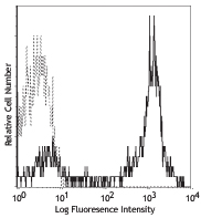
Lymph node cells from a C57BL/6 mouse, at day 12, post-immun... 
Overlay between germinal center (IgD-/Fas+ -
Alexa Fluor® 647 anti-mouse/human Bcl-6

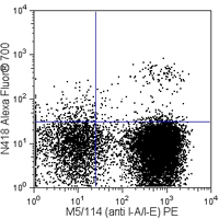
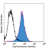
C57BL/6 lymph node cells were immunized with CFA for 12 days... 
-
Alexa Fluor® 594 anti-mouse/human Bcl-6

Human paraffin-embedded tonsil tissue slices were prepared w...


Follow Us