 Login / Register


- Clone
- HIR2 (See other available formats)
- Regulatory Status
- RUO
- Workshop
- VII 70299
- Other Names
- Glycophorin A/B, GPA/GPB, GYPA, GYPB
- Isotype
- Mouse IgG2b, κ
- Ave. Rating
- Submit a Review
- Product Citations
- publications

-

Human red blood cells stained with CD235a/b PE -

IHC staining of purified anti-human CD235ab antibody (clone HIR2) on formalin-fixed paraffin-embedded human spleen tissue. Following antigen retrieval using 0.1 M Tris-buffered saline with Tween 20 (Cat. No. 925501), the tissue was incubated with 1 µg/mL of the primary antibody overnight at 4°C, followed by incubation with 2.5 µg/mL of Alexa Fluor® 647 goat anti-mouse IgG (green) (Cat. No. 405322) for one hour at room temperature. Nuclei were counterstained with DAPI (blue), and the slide was mounted with ProLong™ Gold Antifade Mountant. The image was captured with a 10x objective. Scale bar: 100 µm -

SeqIF™ (sequential immunofluorescence) staining on COMET™ of Purified anti-CD235ab (clone HIR2, yellow) on formalin-fixed paraffin-embedded human pancreatic carcinoma at 0.83 µg/mL. Alexa Fluor™ Plus 647 Goat anti-Mouse IgG antibody (Lunaphore, Cat. No. DR647MS) was used as a secondary antibody. Nuclei were counterstained with DAPI (blue). Tissue underwent an all-in-one dewaxing and antigen retrieval preprocessing.
| Cat # | Size | Price | Quantity Check Availability | Save | ||
|---|---|---|---|---|---|---|
| 306602 | 100 µg | 82€ | ||||
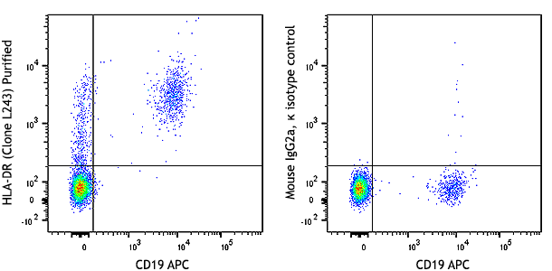
The HIR2 antibody reacts with a common epitope of glycophorin A (CD235a) and glycophorin B (CD235b). Glycophorin A is the major sialoglycoprotein expressed on red blood cell membrane, and erythroid precursors. Glycophorin A shares strong homology with glycophorin B. The HIR2 antibody recognizes human RBCs and erythroid precursors and is useful in erythroid cell development studies. Mature, non-nucleated red blood cells are characteristically glycophorin A positive, but CD45 and CD71 negative.
Product DetailsProduct Details
- Verified Reactivity
- Human
- Reported Reactivity
- African Green, Baboon
- Antibody Type
- Monoclonal
- Host Species
- Mouse
- Formulation
- Phosphate-buffered solution, pH 7.2, containing 0.09% sodium azide.
- Preparation
- The antibody was purified by affinity chromatography.
- Concentration
- 0.5 mg/mL
- Storage & Handling
- The antibody solution should be stored undiluted between 2°C and 8°C.
- Application
-
FC - Quality tested
CyTOF®, IHC-P - Verified
SB - Community verified - Recommended Usage
-
Each lot of this antibody is quality control tested by immunofluorescent staining with flow cytometric analysis. For flow cytometric staining, the suggested use of this reagent is ≤ 0.125 µg per million cells in 100 µL volume. For immunohistochemistry on formalin-fixed paraffin-embedded tissue sections, a concentration range of 1.0 µg/ml is suggested. It is recommended that the reagent be titrated for optimal performance for each application.
- Additional Product Notes
-
For the use of this antibody in spatial biology applications, we have partnered with Lunaphore Technologies for demonstration of our antibodies on the COMET™. The COMET™ platform is an automated, end-to-end spatial biology solution developed for rapid and flexible multiplex tissue profiling. More information on the COMET™ and a complete list of our antibodies that have been demonstrated on the COMET™ can be found here.
-
Application References
(PubMed link indicates BioLegend citation) -
- Mason D, et al. Eds. 2002. Leucocyte Typing VII. Oxford University Press. New York.
- Product Citations
-
- RRID
-
AB_314620 (BioLegend Cat. No. 306602)
Antigen Details
- Structure
- Sialoglycoprotein, 20 kD
- Distribution
-
Erythrocytes
- Cell Type
- Erythrocytes
- Biology Area
- Immunology
- Molecular Family
- CD Molecules
- Antigen References
-
1. Mason D, et al. Eds. 2002. Leucocyte Typing VII. Oxford University Press. New York.
- Gene ID
- 2993 View all products for this Gene ID
- UniProt
- View information about CD235ab on UniProt.org


Related FAQs
- If an antibody clone has been previously successfully used in IBEX in one fluorescent format, will other antibody formats work as well?
-
It’s likely that other fluorophore conjugates to the same antibody clone will also be compatible with IBEX using the same sample fixation procedure. Ultimately a directly conjugated antibody’s utility in fluorescent imaging and IBEX may be specific to the sample and microscope being used in the experiment. Some antibody clone conjugates may perform better than others due to performance differences in non-specific binding, fluorophore brightness, and other biochemical properties unique to that conjugate.
- Will antibodies my lab is already using for fluorescent or chromogenic IHC work in IBEX?
-
Fundamentally, IBEX as a technique that works much in the same way as single antibody panels or single marker IF/IHC. If you’re already successfully using an antibody clone on a sample of interest, it is likely that clone will have utility in IBEX. It is expected some optimization and testing of different antibody fluorophore conjugates will be required to find a suitable format; however, legacy microscopy techniques like chromogenic IHC on fixed or frozen tissue is an excellent place to start looking for useful antibodies.
- Are other fluorophores compatible with IBEX?
-
Over 18 fluorescent formats have been screened for use in IBEX, however, it is likely that other fluorophores are able to be rapidly bleached in IBEX. If a fluorophore format is already suitable for your imaging platform it can be tested for compatibility in IBEX.
- The same antibody works in one tissue type but not another. What is happening?
-
Differences in tissue properties may impact both the ability of an antibody to bind its target specifically and impact the ability of a specific fluorophore conjugate to overcome the background fluorescent signal in a given tissue. Secondary stains, as well as testing multiple fluorescent conjugates of the same clone, may help to troubleshoot challenging targets or tissues. Using a reference control tissue may also give confidence in the specificity of your staining.
- How can I be sure the staining I’m seeing in my tissue is real?
-
In general, best practices for validating an antibody in traditional chromogenic or fluorescent IHC are applicable to IBEX. Please reference the Nature Methods review on antibody based multiplexed imaging for resources on validating antibodies for IBEX.
Other Formats
View All CD235ab Reagents Request Custom Conjugation| Description | Clone | Applications |
|---|---|---|
| APC anti-human CD235ab | HIR2 | FC |
| PE anti-human CD235ab | HIR2 | FC |
| PE/Cyanine5 anti-human CD235ab | HIR2 | FC |
| Purified anti-human CD235ab | HIR2 | FC,CyTOF®,IHC-P,SB |
| FITC anti-human CD235ab | HIR2 | FC |
| Pacific Blue™ anti-human CD235ab | HIR2 | FC |
| PerCP/Cyanine5.5 anti-human CD235ab | HIR2 | FC |
| Purified anti-human CD235ab (Maxpar® Ready) | HIR2 | FC,CyTOF® |
| Biotin anti-human CD235ab | HIR2 | FC |
| PE/Cyanine7 anti-human CD235ab | HIR2 | FC |
| APC/Fire™ 750 anti-human CD235ab | HIR2 | FC |
| TotalSeq™-A0196 anti-human CD235ab | HIR2 | PG |
| TotalSeq™-C0196 anti-human CD235ab | HIR2 | PG |
| TotalSeq™-B0196 anti-human CD235ab | HIR2 | PG |
| TotalSeq™-D0196 anti-human CD235ab | HIR2 | PG |
| TotalSeq™-Bn0196 anti-human CD235ab | HIR2 | SB |
Customers Also Purchased
Compare Data Across All Formats
This data display is provided for general comparisons between formats.
Your actual data may vary due to variations in samples, target cells, instruments and their settings, staining conditions, and other factors.
If you need assistance with selecting the best format contact our expert technical support team.
-
APC anti-human CD235ab

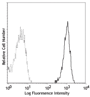
Human red blood cells stained with HIR2 APC -
PE anti-human CD235ab

Human red blood cells stained with CD235a/b PE -
PE/Cyanine5 anti-human CD235ab

Human red blood cells stained with HIR2 PE/Cyanine5 -
Purified anti-human CD235ab
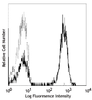
Human red blood cells stained with CD235a/b PE 
IHC staining of purified anti-human CD235ab antibody (clone ... 
SeqIF™ (sequential immunofluorescence) staining on COMET™ of... -
FITC anti-human CD235ab

Human peripheral red blood cells stained with HIR2 FITC -
Pacific Blue™ anti-human CD235ab

Human erythrocytes stained with HIR2 Pacific Blue™ -
PerCP/Cyanine5.5 anti-human CD235ab

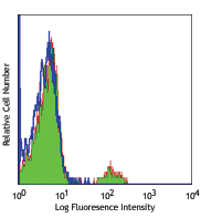
Human red blood cells were stained with anti-human CD235ab ... -
Purified anti-human CD235ab (Maxpar® Ready)
-
Biotin anti-human CD235ab

Human peripheral blood red cells were stained with biotinyla... -
PE/Cyanine7 anti-human CD235ab

Human erythrocytes were first treated with Human TruStain Fc... -
APC/Fire™ 750 anti-human CD235ab

Human erythrocytes were first treated with Human TruStain Fc... -
TotalSeq™-A0196 anti-human CD235ab
-
TotalSeq™-C0196 anti-human CD235ab
-
TotalSeq™-B0196 anti-human CD235ab
-
TotalSeq™-D0196 anti-human CD235ab
-
TotalSeq™-Bn0196 anti-human CD235ab


Follow Us