 Login / Register


- Clone
- 1B7 (See other available formats)
- Regulatory Status
- RUO
- Other Names
- RNA binding protein fox-1 homolog 3, fox-1 homolog C, neuronal nuclei, hexaribonucleotide binding protein 3
- Previously
-
Covance Catalog# SIG-39860
- Isotype
- Mouse IgG2b, κ
- Ave. Rating
- Submit a Review
- Product Citations
- publications

-

IHC staining of purified anti-FOX3 (NeuN) antibody (clone 1B7) on formalin-fixed paraffin-embedded mouse brain tissue. Following antigen retrieval using Sodium Citrate H.I.E.R., the tissue was incubated with 0.5 µg/mL of the primary antibody overnight at 4°C. BioLegend’s Ultra Streptavidin (USA) HRP Detection Kit (Multi-Species, DAB, Cat. No. 929901) was used for detection followed by hematoxylin counterstaining, according to the protocol provided. The image was captured with a 4X objective. Scale bar: 300 µm -

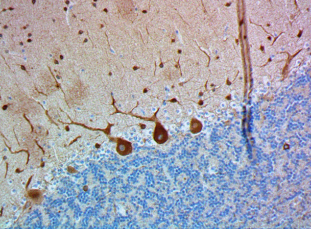
IHC staining of purified anti-FOX3 (NeuN) antibody (clone 1B7) on formalin-fixed paraffin-embedded mouse brain tissue. Following antigen retrieval using Sodium Citrate H.I.E.R., the tissue was incubated with 0.5 µg/mL of the primary antibody overnight at 4°C. BioLegend’s Ultra Streptavidin (USA) HRP Detection Kit (Multi-Species, DAB, Cat. No. 929901) was used for detection followed by hematoxylin counterstaining, according to the protocol provided. The image was captured with a 10X objective. Scale bar: 100 µm -

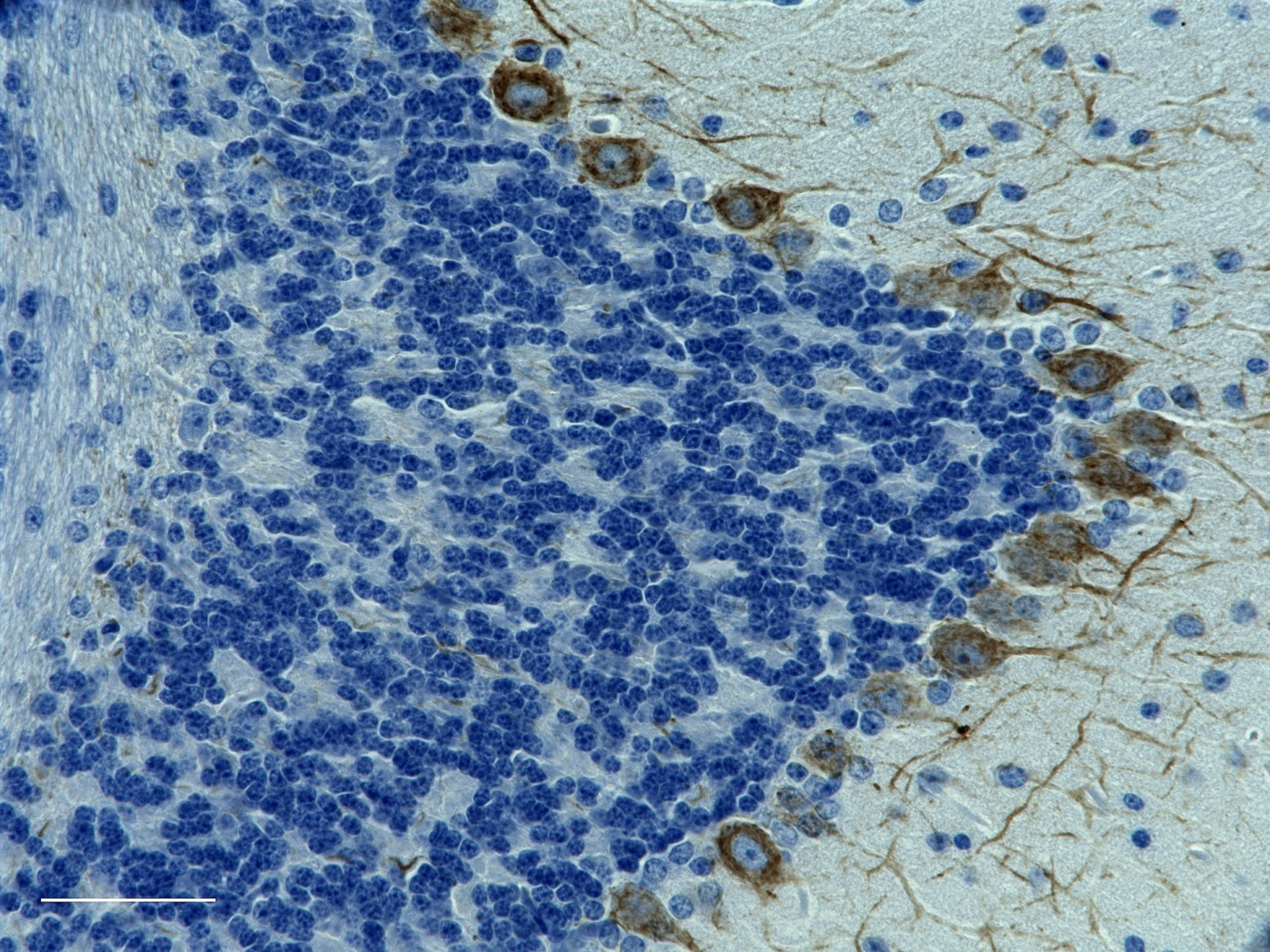
IHC staining of purified anti-FOX3 (NeuN) antibody (clone 1B7) on formalin-fixed paraffin-embedded rat brain tissue. Following antigen retrieval using Sodium Citrate H.I.E.R., the tissue was incubated with 0.5 µg/mL of the primary antibody overnight at 4°C. BioLegend’s Ultra Streptavidin (USA) HRP Detection Kit (Multi-Species, DAB, Cat. No. 929901) was used for detection followed by hematoxylin counterstaining, according to the protocol provided. The image was captured with a 10X objective. Scale bar: 100 µm -

Western blot of purified anti-FOX3 (NeuN) antibody (clone 1B7). Lane 1: Molecular weight marker; Lane 2: 20 µg of human brain lysate; Lane 3: 20 µg of mouse brain lysate; Lane 4: 20 µg of rat brain lysate. The blot was incubated with 0.05 µg/mL of the primary antibody overnight at 4°C, followed by incubation with HRP goat anti-mouse IgG antibody (Cat. No. 405306). Enhanced chemiluminescence was used as the detection system. -

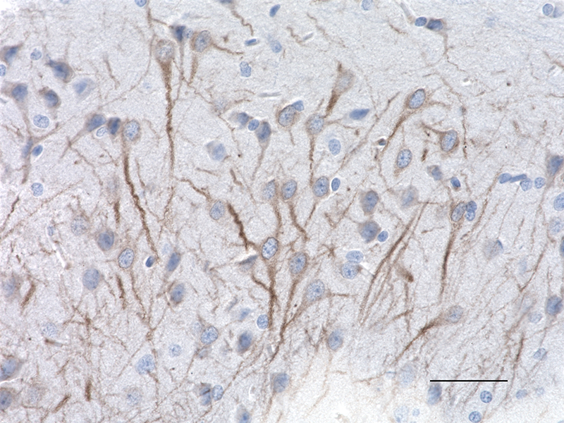
IHC staining of purified anti-FOX3 (NeuN) antibody (clone 1B7) on formalin-fixed paraffin-embedded human brain tissue. Following antigen retrieval using Sodium Citrate H.I.E.R., the tissue was incubated with 1 µg/mL of the primary antibody for 60 minutes at room temperature. BioLegend’s Ultra Streptavidin (USA) HRP Detection Kit (Multi-Species, DAB, Cat. No. 929901) was used for detection followed by hematoxylin counterstaining, according to the protocol provided. The image was captured with a 40X objective. -

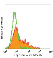
A431 cells (low expression control, open histogram), or A549 cells (positive control, filled histogram) were fixed and permeabilized using True-Nuclear™ Transcription Factor Buffer Set (Cat. No. 424401) and intracellularly stained with purified anti-FOX3 (NeuN) (clone 1B7), or purified mouse IgG2b, κ isotype control (open histogram, dashed line) (representative histogram for both cell lines) (Cat. No. 400302), followed by PE goat anti-mouse IgG (Cat. No. 405307).
| Cat # | Size | Price | Quantity Check Availability | Save | ||
|---|---|---|---|---|---|---|
| 834502 | 25 µL | 90€ | ||||
| 834501 | 100 µL | 191€ | ||||
Fox3 is one of a family of 3 mammalian Fox homologues. Fox was discovered in C. elegans as a gene involved in sex determination, and the name Fox is an acronym of "Feminizing locus on X". The Fox protein and its 3 mammalian homologues are all about 46 kD proteins, each of which includes a central highly conserved RRM type RNA recognition motif, which corresponds to a small ~70 amino acid structure consisting of 4 beta strands and two alpha-helices. Fox3 is a protein which has a function in RNA splicing and is expressed heavily and specifically in neuronal nuclei. The 1B7 antibody reveals strong nuclear and distal cytoplasmic staining for Fox3/NeuN and the complete absence of staining of astrocytes and other kinds of non-neuronal cells. This Fox3/NeuN antibody is therefore an excellent marker of neuronal cells.
Product DetailsProduct Details
- Verified Reactivity
- Human, Mouse, Rat
- Antibody Type
- Monoclonal
- Host Species
- Mouse
- Immunogen
- Antibody was raised against the N-terminal 100 amino acids of human Fox3 as expressed in and purified from E. coli.
- Formulation
- Phosphate-buffered solution + 10mM sodium azide.
- Preparation
- The antibody was purified by affinity chromatography.
- Concentration
- 1 mg/mL
- Storage & Handling
- The antibody solution should be stored undiluted between 2°C and 8°C. Please note the storage condition for this antibody has been changed from -20°C to between 2°C and 8°C. You can also check your vial or your CoA to find the most accurate storage condition for this antibody.
- Application
-
IHC-P - Quality tested
WB, ICFC - Verified
ICC, IHC-F - Reported in the literature, not verified in house
SB - Community verified - Recommended Usage
-
Each lot of this antibody is quality control tested by formalin-fixed paraffin-embedded immunohistochemical staining. For immunohistochemistry, a concentration range of 0.5 - 1.0 µg/mL is suggested. For western blotting, the suggested use of this reagent is 0.01 - 0.05 µg/mL. For intracellular flow cytometric staining, the suggested use of this reagent is ≤ 0.125 µg per million cells in 100 µL volume. It is recommended that the reagent be titrated for optimal performance for each application.
- Application Notes
-
The double band observed in the immunoblot corresponds to the two NeuN isoforms produced by alternative splicing with expected molecular weights of 46 kD and 48 kD.
- Additional Product Notes
-
This product has been verified for IHC-F (Immunohistochemistry - frozen tissue sections) on the NanoString GeoMx® Digital Spatial Profiler. The GeoMx® enables researchers to perform spatial analysis of protein and RNA targets in FFPE and fresh frozen human and mouse samples. For more information about our spatial biology products and the GeoMx® platform, please visit our spatial biology page.
-
Application References
(PubMed link indicates BioLegend citation) -
- Herculano-Houzel S and Lent R. 2005. J Neurosci. 25:2518-21. (ICC)
- Kim KK, et al. 2009. J Biol Chem. 284:31052-31061. (WB, ICC, IHC-P)
- Underwood JG, et al. 2005. Mol Cell Biol. 25:10005-10016. (IHC-F, WB)
- Product Citations
-
- RRID
-
AB_2734601 (BioLegend Cat. No. 834502)
AB_2564991 (BioLegend Cat. No. 834501)
Antigen Details
- Structure
- Expected MW: 46 kD and 48 kD
- Cell Type
- Mature Neurons, Neural Stem Cells
- Biology Area
- Cell Biology, Neuroscience, Neuroscience Cell Markers, Stem Cells
- Molecular Family
- Nuclear Markers
- Gene ID
- 146713 View all products for this Gene ID
- UniProt
- View information about FOX3 on UniProt.org

Related FAQs
- If an antibody clone has been previously successfully used in IBEX in one fluorescent format, will other antibody formats work as well?
-
It’s likely that other fluorophore conjugates to the same antibody clone will also be compatible with IBEX using the same sample fixation procedure. Ultimately a directly conjugated antibody’s utility in fluorescent imaging and IBEX may be specific to the sample and microscope being used in the experiment. Some antibody clone conjugates may perform better than others due to performance differences in non-specific binding, fluorophore brightness, and other biochemical properties unique to that conjugate.
- Will antibodies my lab is already using for fluorescent or chromogenic IHC work in IBEX?
-
Fundamentally, IBEX as a technique that works much in the same way as single antibody panels or single marker IF/IHC. If you’re already successfully using an antibody clone on a sample of interest, it is likely that clone will have utility in IBEX. It is expected some optimization and testing of different antibody fluorophore conjugates will be required to find a suitable format; however, legacy microscopy techniques like chromogenic IHC on fixed or frozen tissue is an excellent place to start looking for useful antibodies.
- Are other fluorophores compatible with IBEX?
-
Over 18 fluorescent formats have been screened for use in IBEX, however, it is likely that other fluorophores are able to be rapidly bleached in IBEX. If a fluorophore format is already suitable for your imaging platform it can be tested for compatibility in IBEX.
- The same antibody works in one tissue type but not another. What is happening?
-
Differences in tissue properties may impact both the ability of an antibody to bind its target specifically and impact the ability of a specific fluorophore conjugate to overcome the background fluorescent signal in a given tissue. Secondary stains, as well as testing multiple fluorescent conjugates of the same clone, may help to troubleshoot challenging targets or tissues. Using a reference control tissue may also give confidence in the specificity of your staining.
- How can I be sure the staining I’m seeing in my tissue is real?
-
In general, best practices for validating an antibody in traditional chromogenic or fluorescent IHC are applicable to IBEX. Please reference the Nature Methods review on antibody based multiplexed imaging for resources on validating antibodies for IBEX.
Other Formats
View All FOX3 Reagents Request Custom Conjugation| Description | Clone | Applications |
|---|---|---|
| Purified anti-FOX3 (NeuN) | 1B7 | IHC-P,WB,ICFC,ICC,IHC-F,SB |
Customers Also Purchased
Compare Data Across All Formats
This data display is provided for general comparisons between formats.
Your actual data may vary due to variations in samples, target cells, instruments and their settings, staining conditions, and other factors.
If you need assistance with selecting the best format contact our expert technical support team.
-
Purified anti-FOX3 (NeuN)

IHC staining of purified anti-FOX3 (NeuN) antibody (clone 1B... 
IHC staining of purified anti-FOX3 (NeuN) antibody (clone 1B... 
A431 cells (low expression control, open histogram), or A549... 
IHC staining of purified anti-FOX3 (NeuN) antibody (clone 1B... 
IHC staining of purified anti-FOX3 (NeuN) antibody (clone 1B... 
Western blot of purified anti-FOX3 (NeuN) antibody (clone 1B...


Follow Us