 Login / Register


- Clone
- 6A7 (See other available formats)
- Regulatory Status
- RUO
- Other Names
- BCL2 associated X protein, apoptosis regulator Bax
- Isotype
- Mouse IgG1, κ
- Ave. Rating
- Submit a Review
- Product Citations
- publications

-

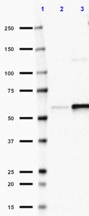
Immunoprecipitation/Western blot analysis of extracts from Daudi cells(lane 1 and lane 2) and THP-1 cells (lane 4 and lane5). Lane 4 contains THP-1 lysate input (5%). Lane 1 and lane 4 were immunoprecipitated with mouse IgG1 isotype control antibodies and lane 2 and lane 5 were immunoprecipitated with BAX antibody (6A7). Western blot analysis was performed using rabbit anti-BAX antibody (poly6251).
| Cat # | Size | Price | Quantity Check Availability | Save | ||
|---|---|---|---|---|---|---|
| 633801 | 25 µg | 81€ | ||||
| 633802 | 100 µg | 177€ | ||||
Bax is a 21 kD pro-apoptotic protein known to regulate apoptosis. Bax is found in the cytoplasm, mitochondria, and nucleus and is highly expressed in hematopoietic stem cells, the ovary, and in the lymph node. Bax binds the anti-apoptotic protein Bcl-2 as a heterodimer or forms homodimers. The relative levels of pro-apoptotic proteins such as Bax and anti-apoptotic proteins such as Bcl-2 determines whether cell death will occur following an apoptotic stimulus. Bax accelerates the opening of mitochondrial VDAC altering membrane potential and allowing cytochrome c to pass out of the mitochondria into the cytosol to initiate downstream caspase activation. p53 can transcriptionally activate the Bax gene to induce apoptosis. Bax has been shown to be mutated in some human cancers. Clone 2D2 has been shown to be useful for western blotting, immunoprecipitation, immunofluorescence and immunohistochemistry of the human Bax protein.This antibody does not cross-react with Bcl-2 or Bcl-XL proteins.
Product DetailsProduct Details
- Verified Reactivity
- Human, Mouse, Rat, Monkey
- Antibody Type
- Monoclonal
- Host Species
- Mouse
- Immunogen
- Amino acid:12-24 common to human, mouse, and rat Bax protein
- Formulation
- This antibody is provided in phosphate-buffered solution, pH 7.2, containing 0.09% sodium azide at 0.5 mg/ml.
- Preparation
- The antibody was purified by affinity chromatography.
- Concentration
- 0.5 mg/ml
- Storage & Handling
- Upon receipt, store undiluted at between 2°C and 8°C.
- Application
-
IP - Quality tested
- Recommended Usage
-
Each lot of this antibody is quality control tested by immunoprecipitation. This antibody can be used at 2-4 µg /1 x107 cell equivalents for immunoprecipitation. It is recommended that the reagent be titrated for optimal performance for each application.
-
Application References
(PubMed link indicates BioLegend citation) -
- Hsu YT, et al. 1997. J. Biol. Chem. 272:13829.
- Liu G, et al. 2013. J. Immunol. 191:2680. PubMed
- Product Citations
-
- RRID
-
AB_2227699 (BioLegend Cat. No. 633801)
AB_2061543 (BioLegend Cat. No. 633802)
Antigen Details
- Structure
- Forms homodimers and heterodimers with Bcl-2, approximately 21 kD
- Distribution
- Cytoplasm, mitochondria, nucleus. Expressed in hematopoietic stem cells, ovary, lymph node
- Function
- Induces cell death through the process of apoptosis by acting on mitochondria.
- Interaction
- 14-3-3 theta, VDAC1, Bcl-2, solute carrier family 25, mitochondrial carrier member 4
- Cell Type
- Hematopoietic stem and progenitors
- Biology Area
- Apoptosis/Tumor Suppressors/Cell Death, Cell Biology, Immunology, Neuroscience
- Antigen References
-
1. LeBlanc H, et al. 2002. Nat. Med. 8:274.
2. Marzo I, et al. 1998. Science 281:2027.
3. Miyashita T et al. 1995. Cell 80:293.
4. Oltvai ZN, et al. 1993. Cell 74:609. - Gene ID
- 581 View all products for this Gene ID
- UniProt
- View information about Bax on UniProt.org

Related FAQs
Other Formats
View All Bax Reagents Request Custom Conjugation| Description | Clone | Applications |
|---|---|---|
| Purified anti-Bax | 6A7 | IP |
Customers Also Purchased
Compare Data Across All Formats
This data display is provided for general comparisons between formats.
Your actual data may vary due to variations in samples, target cells, instruments and their settings, staining conditions, and other factors.
If you need assistance with selecting the best format contact our expert technical support team.
-
Purified anti-Bax

Immunoprecipitation/Western blot analysis of extracts from D...


Follow Us