 Login / Register


- Clone
- H037G3 (See other available formats)
- Regulatory Status
- RUO
- Other Names
- Macrophage galactose/Nacetylgalactosamine (GalNAc) specific lectin, MGL, DCASGPR, HML CLECSF13, CLECSF14, HML2
- Isotype
- Mouse IgG2a, κ
- Ave. Rating
- Submit a Review
- Product Citations
- publications

-

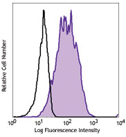
Human monocyte-derived dendritic cells were stained with CD301 (clone H037G3) PE (filled histogram) or mouse IgG2a, κ PE isotype control (open histogram).
| Cat # | Size | Price | Quantity Check Availability | Save | ||
|---|---|---|---|---|---|---|
| 354703 | 25 tests | 123€ | ||||
| 354704 | 100 tests | 288€ | ||||
CD301, also known as CLEC10A, macrophage galactose/N-acetylgalactosamine (GalNAc) specific lectin (MGL), DCASGPR, and HML, is a 40 kD type II transmembrane glycoprotein, which belongs to the C-type lectin family. Human CD301 consists of a 39 amino acid (aa) cytoplasmic region, a 21 aa transmembrane segment, and a 256 aa extracellular domain (ECD) with one carbohydrate recognition domain (CRD) and a neck region. CD301 is expressed on immature myleloid dendritic cells and alternatively activated (tolerogenic) macrophages. The expression level is upregulated by immunosuppressant dexamethasone. Human CD301 has an exclusive specificity for rare terminal GalNAc structures, which are revealed on the tumor-associated mucin MUC1 and CD45 (RA, RB, and RC but not RO isoforms). This interaction inhibits effector T cell activation and induces their apoptosis. CD301 also binds the GP envelope glycoprotein on Marburg and Ebola viruses and enhances viral entry and infectivity.
Product DetailsProduct Details
- Verified Reactivity
- Human
- Reported Reactivity
- African Green, Baboon, Cynomolgus
- Antibody Type
- Monoclonal
- Host Species
- Mouse
- Immunogen
- Recombinant CD301 (extracellular domain)
- Formulation
- Phosphate-buffered solution, pH 7.2, containing 0.09% sodium azide and BSA (origin USA)
- Preparation
- The antibody was purified by affinity chromatography and conjugated with PE under optimal conditions.
- Concentration
- Lot-specific (to obtain lot-specific concentration and expiration, please enter the lot number in our Certificate of Analysis online tool.)
- Storage & Handling
- The antibody solution should be stored undiluted between 2°C and 8°C, and protected from prolonged exposure to light. Do not freeze.
- Application
-
FC - Quality tested
- Recommended Usage
-
Each lot of this antibody is quality control tested by immunofluorescent staining with flow cytometric analysis. For flow cytometric staining, the suggested use of this reagent is 5 µl per million cells in 100 µl staining volume or 5 µl per 100 µl of whole blood.
- Excitation Laser
-
Blue Laser (488 nm)
Green Laser (532 nm)/Yellow-Green Laser (561 nm)
- Application Notes
-
H037G3 antibody binding is divalent cation dependent.
- Product Citations
-
- RRID
-
AB_11219202 (BioLegend Cat. No. 354703)
AB_11219002 (BioLegend Cat. No. 354704)
Antigen Details
- Structure
- 40 kD type II transmembrane glycoprotein, consists of a 39 amino acid (aa) cytoplasmic region, a 21 aa transmembrane segment and a 256 aa extracellular domain (ECD) with one carbohydrate recognition domain (CRD) and a neck region
- Distribution
-
Immature myeloid dendritic cells, activated macrophages
- Function
- Cell adhesion, cell-cell signaling, glycoprotein turnover, inflammation, immune response
- Interaction
- Carbohydrate determinants, GP envelope glycoprotein on Marburg and Ebola viruses
- Ligand/Receptor
- Glycans containing terminal GalNAc moieties
- Cell Type
- Dendritic cells, Macrophages
- Biology Area
- Cell Adhesion, Cell Biology, Immunology, Innate Immunity, Signal Transduction
- Molecular Family
- Adhesion Molecules, CD Molecules
- Antigen References
-
1. Van Vliet SJ, et al. 2006. Nat. Immunol. 7:1200.
2. Higashi N, et al. 2002. J. Biol. Chem. 277:20686.
3. Tsuiji M, et al. 2002. J. Biol. Chem. 277:28892.
4. Valladeau J, et al. 2001. J. Immunol. 167:5767.
5. Suzuki N, et al. 1996. J. Immunol. 156:128. - Gene ID
- 10462 View all products for this Gene ID
- UniProt
- View information about CD301 on UniProt.org
Related Pages & Pathways
Pathways


Related FAQs
- What type of PE do you use in your conjugates?
- We use R-PE in our conjugates.
Other Formats
View All CD301 Reagents Request Custom Conjugation| Description | Clone | Applications |
|---|---|---|
| Purified anti-human CD301 (CLEC10A) | H037G3 | FC |
| PE anti-human CD301 (CLEC10A) | H037G3 | FC |
| APC anti-human CD301 (CLEC10A) | H037G3 | FC |
| TotalSeq™-A0401 anti-human CD301 (CLEC10A) | H037G3 | PG |
Customers Also Purchased
Compare Data Across All Formats
This data display is provided for general comparisons between formats.
Your actual data may vary due to variations in samples, target cells, instruments and their settings, staining conditions, and other factors.
If you need assistance with selecting the best format contact our expert technical support team.
-
Purified anti-human CD301 (CLEC10A)

Human monocyte-derived dendritic cells were stained with pur... -
PE anti-human CD301 (CLEC10A)

Human monocyte-derived dendritic cells were stained with CD3... -
APC anti-human CD301 (CLEC10A)

Human monocyte-derived dendritic cells were stained with CD3... -
TotalSeq™-A0401 anti-human CD301 (CLEC10A)


Follow Us