 Login / Register


- Clone
- HNK-1 (See other available formats)
- Regulatory Status
- RUO
- Other Names
- HNK-1, NK-1, Leu-7
- Isotype
- Mouse IgM, κ
- Ave. Rating
- Submit a Review
- Product Citations
- publications

-

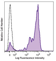
Human peripheral blood lymphocytes were stained with CD8 APC and CD57 (clone HNK-1) Pacific Blue™ (top) or mouse IgM Pacific Blue™ isotype control (bottom). -

| Cat # | Size | Price | Quantity Check Availability | Save | ||
|---|---|---|---|---|---|---|
| 359607 | 25 tests | 100€ | ||||
| 359608 | 100 tests | 249€ | ||||
CD57, also known as HNK-1, NK-1, and Leu-7 is a 100-115 kD oligosaccharide antigenic determinant expressed on a variety of proteins, lipids, and chondroitin sulfate proteoglycans. CD57 is expressed on a subset of peripheral blood lymphocytes, including NK cells and CD8+ T cells, and is also expressed on neural cells and striated muscle. CD57 is not expressed on red blood cells, granulocytes, monocytes, or platelets. While the function of CD57 is unknown, binding to L-selectin, P-selectin, and a fragment of laminin suggests that CD57 may be involved in cell-matrix interactions. CD57 is increased in some disease states associated with CD4/CD8 imbalances (AIDS, autoimmune disease, viral infections, and allograft transplants).
Product DetailsProduct Details
- Verified Reactivity
- Human
- Antibody Type
- Monoclonal
- Host Species
- Mouse
- Immunogen
- Membrane extract of human lymphoblastoid cell line HSB-2.
- Formulation
- Phosphate-buffered solution, pH 7.2, containing 0.09% sodium azide and BSA (origin USA)
- Preparation
- The antibody was purified by affinity chromatography and conjugated with Pacific Blue™ under optimal conditions.
- Concentration
- Lot-specific (to obtain lot-specific concentration and expiration, please enter the lot number in our Certificate of Analysis online tool.)
- Storage & Handling
- The antibody solution should be stored undiluted between 2°C and 8°C, and protected from prolonged exposure to light. Do not freeze.
- Application
-
FC - Quality tested
- Recommended Usage
-
Each lot of this antibody is quality control tested by immunofluorescent staining with flow cytometric analysis. For flow cytometric staining, the suggested use of this reagent is 5 µl per million cells in 100 µl staining volume or 5 µl per 100 µl of whole blood.
* Pacific Blue™ has a maximum emission of 455 nm when it is excited at 405 nm. Prior to using Pacific Blue™ conjugate for flow cytometric analysis, please verify your flow cytometer's capability of exciting and detecting the fluorochrome.
Alexa Fluor® and Pacific Blue™ are trademarks of Life Technologies Corporation.
View full statement regarding label licenses - Excitation Laser
-
Violet Laser (405 nm)
- Application Notes
-
Additional reported applications for the relevant formats include: Western blotting1.
-
Application References
(PubMed link indicates BioLegend citation) -
- Yoshihara Y, et al. 1991. J. Cell Biol. 115:731. (WB)
- Abo T, et al. 1981. J. Immunol. 127:1024.
- Abo T, et al. 1982. J. Immunol. 129:1752.
- Abo T, et al. 1982. J. Immunol. 129:1758.
- RRID
-
AB_2562458 (BioLegend Cat. No. 359607)
AB_2562459 (BioLegend Cat. No. 359608)
Antigen Details
- Structure
- Oligosaccharide antigenic determinant present on a variety of proteins, lipids, and chrondroitin sulfate proteoglycans, 100-115 kD. The antigen is conserved across species.
- Distribution
-
CD57 antigen is expressed on a subset of peripheral blood lymphocytes including a subset of NK cells and a subset of CD8+ T cells. Also expressed on neural cells and striated muscle.
- Function
- Unknown function, may be involved in cell-matrix interactions.
- Ligand/Receptor
- Binds to L-selectin and P-selectin in a calcium-dependent manner, also binds to second globular domain of E8 laminin fragment.
- Cell Type
- Lymphocytes, NK cells, T cells
- Biology Area
- Costimulatory Molecules, Immunology
- Molecular Family
- CD Molecules
- Antigen References
-
1. Schubert J, et al. 1989. In Leucocyte Typing IV (Knapp W, ed) Oxford University Press Oxford pp 711-714.
2. Palmer BE, et al. 2005. J. Immunol. 175:8415.
3. Schachner M, et al. 1995. Trends Neurosci. 18:183.
4. Wood KL, et al. 2005. Clin. Immunol. 117:294. - Gene ID
- 27087 View all products for this Gene ID
- UniProt
- View information about CD57 on UniProt.org
Related Pages & Pathways
Pathways


Related FAQs
Other Formats
View All CD57 Reagents Request Custom Conjugation| Description | Clone | Applications |
|---|---|---|
| Purified anti-human CD57 | HNK-1 | FC,IHC-P,WB |
| FITC anti-human CD57 | HNK-1 | FC,SB |
| Pacific Blue™ anti-human CD57 | HNK-1 | FC |
| APC anti-human CD57 | HNK-1 | FC |
| PE anti-human CD57 | HNK-1 | FC,SB |
| Alexa Fluor® 647 anti-human CD57 | HNK-1 | FC,IHC-P,SB |
| Biotin anti-human CD57 | HNK-1 | FC |
| PE/Dazzle™ 594 anti-human CD57 | HNK-1 | FC |
| PerCP/Cyanine5.5 anti-human CD57 | HNK-1 | FC |
| PE/Cyanine7 anti-human CD57 | HNK-1 | FC |
| Alexa Fluor® 594 anti-human CD57 | HNK-1 | IHC-P,SB |
| Spark Blue™ 515 anti-human CD57 | HNK-1 | FC |
Customers Also Purchased

Compare Data Across All Formats
This data display is provided for general comparisons between formats.
Your actual data may vary due to variations in samples, target cells, instruments and their settings, staining conditions, and other factors.
If you need assistance with selecting the best format contact our expert technical support team.
-
Purified anti-human CD57

Human peripheral blood lymphocytes were stained with CD8 APC... 

Human paraffin-embedded cerebellum tissue slices were prepar... -
FITC anti-human CD57

Human peripheral blood lymphocytes were stained with CD8 APC... 
-
Pacific Blue™ anti-human CD57

Human peripheral blood lymphocytes were stained with CD8 APC... 
-
APC anti-human CD57

Human peripheral blood lymphocytes were stained with CD8 FIT... 
-
PE anti-human CD57

Human peripheral blood lymphocytes were stained with CD8 FIT... 
-
Alexa Fluor® 647 anti-human CD57


Human paraffin-embedded cerebellum tissue slices were prepar... -
Biotin anti-human CD57

Human peripheral blood lymphocytes were stained with CD8 FIT... 
-
PE/Dazzle™ 594 anti-human CD57

Human peripheral blood lymphocytes were stained with CD8 APC... -
PerCP/Cyanine5.5 anti-human CD57

Human peripheral blood lymphocytes were stained with CD8 APC... -
PE/Cyanine7 anti-human CD57

Human peripheral blood lymphocytes were stained with CD8 APC... -
Alexa Fluor® 594 anti-human CD57

Human paraffin-embedded cerebellum tissue slices were prepar... 
Human paraffin-embedded cerebellum tissue slices were prepar... -
Spark Blue™ 515 anti-human CD57

Human peripheral blood cells were surface stained with anti-...


Follow Us