 Login / Register


- Clone
- GoH3 (See other available formats)
- Regulatory Status
- RUO
- Workshop
- HCDM listed
- Other Names
- VLA-6 α chain, α6 integrin, integrin α6, ITGA6
- Isotype
- Rat IgG2a, κ
- Ave. Rating
- Submit a Review
- Product Citations
- publications

-

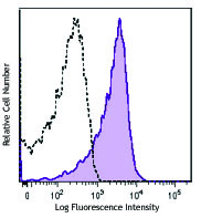
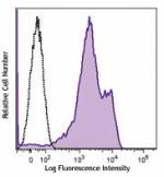
Human peripheral blood lymphocytes were stained with anti-human/mouse CD49f (clone GoH3) Brilliant Violet 421™ (filled histogram) or rat IgG2a, κ Brilliant Violet 421™ isotype control (open histogram). -

Human paraffin-embedded placenta tissue slices were prepared with a standard protocol of deparaffination and rehydration. Antigen retrieval was done with Citrate-Buffered 1X pH 6.0 at 95°C for 40 minutes. Tissue was washed with PBS/0.05% Tween 20 twice for five minutes, permeabilized with 0.5% Tween 20 in PBS and blocked with 5% FBS and 0.2% gelatin for 30 minutes. Then, the tissue was stained with 10 µg/ml of Brilliant Violet 421™ anti-human/mouse CD49f (clone GoH3) antibody (blue) overnight at 4°C. Nuclei were counter-stained with Helix Green (green). The image was scanned with a 10X objective and stitched with MetaMorph® software.
| Cat # | Size | Price | Quantity Check Availability | Save | ||
|---|---|---|---|---|---|---|
| 313623 | 25 tests | 159€ | ||||
| 313624 | 100 tests | 322€ | ||||
CD49f is a 120 kD integrin family member also known as VLA-6 α chain and α6 integrin subunit. CD49f associates with either integrin β1 (CD29) or integrin β4 (CD104) to form receptors (VLA-6 or α6β4 complex) for laminin and kalinin. CD49f is expressed on platelets, monocytes, T cells, placental trophoblasts, and epithelial and endothelial cells. CD49f is involved in adhesion and can act as a co-stimulatory molecule for T cell activation and proliferation.
Product DetailsProduct Details
- Verified Reactivity
- Human, Cynomolgus, Rhesus
- Reported Reactivity
- African Green, Mouse, Baboon, Capuchin Monkey, Cat, Cow, Chimpanzee, Cynomolgus, Dog, Horse, Rabbit, Sheep, Pig
- Antibody Type
- Monoclonal
- Host Species
- Rat
- Immunogen
- Mouse mammary tumor cells
- Formulation
- Phosphate-buffered solution, pH 7.2, containing 0.09% sodium azide and BSA (origin USA).
- Preparation
- The antibody was purified by affinity chromatography and conjugated with Brilliant Violet 421™ under optimal conditions.
- Concentration
- Lot-specific (to obtain lot-specific concentration and expiration, please enter the lot number in our Certificate of Analysis online tool.)
- Storage & Handling
- The antibody solution should be stored undiluted between 2°C and 8°C, and protected from prolonged exposure to light. Do not freeze.
- Application
-
FC - Quality tested
IHC-P - Verified - Recommended Usage
-
Each lot of this antibody is quality control tested by immunofluorescent staining with flow cytometric analysis. For flow cytometric staining, the suggested use of this reagent is 5 µl per million cells in 100 µl staining volume or 5 µl per 100 µl of whole blood. For immunohistochemistry, a concentration range of 5.0 - 10.0 µg/ml is suggested. It is recommended that the reagent be titrated for optimal performance for each application.
Brilliant Violet 421™ excites at 405 nm and emits at 421 nm. The standard bandpass filter 450/50 nm is recommended for detection. Brilliant Violet 421™ is a trademark of Sirigen Group Ltd.
Learn more about Brilliant Violet™.
This product is subject to proprietary rights of Sirigen Inc. and is made and sold under license from Sirigen Inc. The purchase of this product conveys to the buyer a non-transferable right to use the purchased product for research purposes only. This product may not be resold or incorporated in any manner into another product for resale. Any use for therapeutics or diagnostics is strictly prohibited. This product is covered by U.S. Patent(s), pending patent applications and foreign equivalents. - Excitation Laser
-
Violet Laser (405 nm)
- Application Notes
-
Additional reported applications (for the relevant formats) include: immunoprecipitation1,5, in vitro and in vivo blocking of cell binding to laminin and blocking the function of integrin a61,4, and immunohistochemistry of acetone-fixed frozen sections2,3,5. The GoH3 antibody has been reported to block laminin binding in vitro and to block integrin a6 function in vivo.
-
Application References
(PubMed link indicates BioLegend citation) -
- Georas SN, et al. 1993. Blood 82:2872. (IP, Block)
- Honda T, et al. 1995. J. Clin. Endocrinol. Metab. 80:2899. (IHC)
- Sonnenberg A, et al. 1986. J. Histochem. Cytochem. 34:1037. (IHC)
- Nakamura K, et al. 1997 Biochem. Biophys. Res. Commun. 235:524. (Block)
- Sonnenberg A, et al. 1987 J. Biol. Chem. 262:10376. (IP, IHC)
- Deregibus MC, et al. 2007. Blood doi:10.1182/blood-2007-03-078709.
- Horwitz KB, et al. 2008. Proc Natl Acad Sci USA. 105:5774. PubMed
- Nardella C, et al. 2009. Sci Signal. 2:55. PubMed
- Xu T, et al. 2010. Mol Cancer Ther. 9:438. PubMed
- Stepp MA, et al. 2007. J Cell Sci. 120:2851. PubMed
- Jo M, et al. 2010. Cancer Res. 70:8948. PubMed
- Yoshino N, et al. 2000. Exp. Anim. (Tokyo) 49:97. (FC)
- Grange C, et al. 2011. Cancer Res. 71:5346. PubMed
- Lai KP, et al. 2012. Mol Endocrinol. 26:52. PubMed
- Oeztuerk-Winder F, et al. 2012. EMBO J. 31:3431. (FC) PubMed
- Product Citations
-
- RRID
-
AB_2562243 (BioLegend Cat. No. 313623)
AB_2562244 (BioLegend Cat. No. 313624)
Antigen Details
- Structure
- Integrin family, associates with β1 or β4, 120 kD
- Distribution
-
Platelets, monocytes, T cells, placental trophoblasts, epithelial and endothelial cells
- Function
- Adhesion, receptor for laminin and kalinin; laminin binding to VLA-6 induces T cell co-stimulation for proliferation and activation
- Ligand/Receptor
- With integrin β1 (CD29) forms VLA-6, with integrin β4 (CD104) forms a6β4 integrin; laminin and kalinin are ligands for these receptors
- Cell Type
- Embryonic Stem Cells, Endothelial cells, Epithelial cells, Monocytes, Platelets, T cells
- Biology Area
- Cell Adhesion, Cell Biology, Immunology, Innate Immunity, Stem Cells
- Molecular Family
- Adhesion Molecules, CD Molecules
- Antigen References
-
1. Sonnenberg A, et al. 1990. J. Cell Biol. 110:2145.
2. Sonnenberg A, et al. 1990. J. Cell. Sci. 96:207.
3. Aumailley M, et al. 1990. Exp. Cell Res. 188:55.
4. Niessen CM, et al. 1994. Exp. Cell Res. 211:360. - Gene ID
- 16403 View all products for this Gene ID 3655 View all products for this Gene ID
- UniProt
- View information about CD49f on UniProt.org
Related Pages & Pathways
Pathways


Related FAQs
- What is the F/P ratio range of our BV421™ format antibody reagents?
-
It is lot-specific. On average it ranges between 2-4.
Other Formats
View All CD49f Reagents Request Custom Conjugation| Description | Clone | Applications |
|---|---|---|
| Purified anti-human/mouse CD49f | GoH3 | FC,ICC,IHC-F,IP,Block |
| Biotin anti-human/mouse CD49f | GoH3 | FC |
| FITC anti-human/mouse CD49f | GoH3 | FC |
| Alexa Fluor® 488 anti-human/mouse CD49f | GoH3 | FC |
| Alexa Fluor® 647 anti-human/mouse CD49f | GoH3 | FC |
| PE anti-human/mouse CD49f | GoH3 | FC |
| APC anti-human/mouse CD49f | GoH3 | FC |
| PerCP/Cyanine5.5 anti-human/mouse CD49f | GoH3 | FC |
| Pacific Blue™ anti-human/mouse CD49f | GoH3 | FC |
| PE/Cyanine7 anti-human/mouse CD49f | GoH3 | FC |
| Brilliant Violet 421™ anti-human/mouse CD49f | GoH3 | FC,IHC-P |
| PE/Dazzle™ 594 anti-human/mouse CD49f | GoH3 | FC |
| APC/Cyanine7 anti-human/mouse CD49f | GoH3 | FC |
| APC/Fire™ 750 anti-human/mouse CD49f | GoH3 | FC |
| TotalSeq™-A0070 anti-human/mouse CD49f | GoH3 | PG |
| TotalSeq™-C0070 anti-human/mouse CD49f | GoH3 | PG |
| Ultra-LEAF™ Purified anti-human/mouse CD49f | GoH3 | FC,ICC,IHC-F,IP,Block |
| TotalSeq™-B0070 anti-human/mouse CD49f | GoH3 | PG |
| TotalSeq™-D0070 anti-human/mouse CD49f | GoH3 | PG |
Customers Also Purchased
Compare Data Across All Formats
This data display is provided for general comparisons between formats.
Your actual data may vary due to variations in samples, target cells, instruments and their settings, staining conditions, and other factors.
If you need assistance with selecting the best format contact our expert technical support team.
-
Purified anti-human/mouse CD49f

Human peripheral blood lymphocytes stained with purified GoH... 
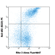
MDA-MB-231 breast cancer cells were stained with anti-CD49f ... -
Biotin anti-human/mouse CD49f

C57BL/6 mouse splenocytes stained with biotinylated GoH3, fo... -
FITC anti-human/mouse CD49f

Human peripheral blood lymphocytes stained with GoH3 FITC -
Alexa Fluor® 488 anti-human/mouse CD49f

Human peripheral blood lymphocytes were stained with anti-hu... -
Alexa Fluor® 647 anti-human/mouse CD49f

Human peripheral blood lymphocytes stained with GoH3 Alexa F... -
PE anti-human/mouse CD49f

Human peripheral blood lymphocytes stained with GoH3 PE -
APC anti-human/mouse CD49f

Human peripheral blood lymphocytes were stained with anti-hu... -
PerCP/Cyanine5.5 anti-human/mouse CD49f

Human peripheral blood lymphocytes stained with GoH3 PerCP/C... -
Pacific Blue™ anti-human/mouse CD49f

Human peripheral blood lymphocytes stained with GoH3 Pacific... -
PE/Cyanine7 anti-human/mouse CD49f

Human peripheral blood lymphocytes were stained with anti-hu... -
Brilliant Violet 421™ anti-human/mouse CD49f
Human peripheral blood lymphocytes were stained with anti-hu... 
Human paraffin-embedded placenta tissue slices were prepared... -
PE/Dazzle™ 594 anti-human/mouse CD49f

Human peripheral blood lymphocytes were stained with anti-hu... -
APC/Cyanine7 anti-human/mouse CD49f

Human peripheral blood lymphocytes were stained with CD49f (... -
APC/Fire™ 750 anti-human/mouse CD49f

Human peripheral blood lymphocytes were stained with anti-hu... -
TotalSeq™-A0070 anti-human/mouse CD49f
-
TotalSeq™-C0070 anti-human/mouse CD49f
-
Ultra-LEAF™ Purified anti-human/mouse CD49f
Human peripheral blood lymphocytes stained with purified GoH...
MDA-MB-231 breast cancer cells were stained with anti-CD49f ... -
TotalSeq™-B0070 anti-human/mouse CD49f
-
TotalSeq™-D0070 anti-human/mouse CD49f


Follow Us