 Login / Register


- Clone
- 17A2 (See other available formats)
- Regulatory Status
- RUO
- Other Names
- T cell antigen receptor complex, T3
- Isotype
- Rat IgG2b, κ
- Ave. Rating
- Submit a Review
- Product Citations
- publications

-

C57BL/6 mouse frozen spleen section was fixed with 4% paraformaldehyde (PFA) for 10 minutes at room temperature and blocked with 5% FBS plus 5% rat serum for 1 hour at room temperature. Then the section was stained with 2.5 µg/mL of CD3 (clone 17A2) Alexa Fluor® 594 (red) and 2.5 µg/mL of B220 (clone RA3-6B2) Alexa Fluor® 488 (green) overnight at 4°C. The image was captured by 10X objective. -

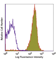
C57BL/6 mouse splenocytes were stained with CD3 (clone 17A2) Alexa Fluor® 594 (filled histogram). The data was acquired by BD LSRFortessa™ cell analyzer equipped with Yellow-Green Laser (561 nm). -
Formalin-fixed, 300 micron-thick mouse spleen section was blocked, permeabilized and stained overnight with CD3 (clone 17A2) Alexa Fluor® 594 (red), CD169 (Siglec-1)(clone 3D6.112) Alexa Fluor® 647 (green) both at 5 µg/mL, optically cleared, then analyzed at 225 μm imaging depth on a confocal microscope. Watch the video. -

Paraformaldehyde-fixed (4%), 500 μm-thick mouse spleen section was processed according to the Ce3DTM Tissue Clearing Kit protocol (cat. no. 47701). The section was costained with anti-mouse CD68 Antibody (clone FA-11) Alexa Fluor® 488 at 5 µg/mL (green), anti-mouse CD3 Antibody (clone 17A2) Alexa Fluor® 594 at 5 µg/mL (yellow), and anti-mouse IgD Antibody (clone 11-26c.2a) Alexa Fluor® 647 at 5 µg/mL (magenta). The section was then optically cleared and mounted in a sample chamber. The image was captured with a 10X objective using Zeiss 780 confocal microscope and processed by Imaris image analysis software.
Watch the video. -

Mice were injected subcutaneously with sheep red blood cells in a volume of 25 l per site on days 0 and 4 and harvested on day 11. Confocal image of C57BL/6 mouse lymph node acquired using the IBEX method of highly multiplexed antibody-based imaging: CD3 (cyan) in Cycle 1 and MHCII (blue) in Cycle 10. Tissues were prepared using ~1% (vol/vol) formaldehyde and a detergent. Following fixation, samples are immersed in 30% (wt/vol) sucrose for cryoprotection. Images are courtesy of Drs. Andrea J. Radtke and Ronald N. Germain of the Center for Advanced Tissue Imaging (CAT-I) in the National Institute of Allergy and Infectious Diseases (NIAID, NIH).

| Cat # | Size | Price | Quantity Check Availability | Save | ||
|---|---|---|---|---|---|---|
| 100240 | 100 µg | 203€ | ||||
CD3, also known as T3, is a member of the Ig superfamily and primarily expressed on T cells, NK-T cells, and at different levels on thymocytes during T cell differentiation. CD3 is composed of CD3ε, δ, γ and ζ chains. It forms a TCR complex by associating with TCR α/β or γ/δ chains. CD3 plays a critical role in TCR signal transduction, T cell activation, and antigen recognition by binding the peptide/MHC antigen complex
Product DetailsProduct Details
- Verified Reactivity
- Mouse
- Antibody Type
- Monoclonal
- Host Species
- Rat
- Immunogen
- γδTCR-positive T-T hybridoma D1
- Formulation
- Phosphate-buffered solution, pH 7.2, containing 0.09% sodium azide.
- Preparation
- The antibody was purified by affinity chromatography and conjugated with Alexa Fluor® 594 under optimal conditions.
- Concentration
- 0.5 mg/mL
- Storage & Handling
- The antibody solution should be stored undiluted between 2°C and 8°C, and protected from prolonged exposure to light. Do not freeze.
- Application
-
IHC-F - Quality tested
FC, 3D IHC - Verified
SB - Reported in the literature, not verified in house
- Recommended Usage
-
Each lot of this antibody is quality control tested by immunohistochemical staining on frozen tissue sections. For immunohistochemistry, a concentration range of 1.0 - 5.0 µg/mL is suggested. For flow cytometric staining, the suggested use of this reagent is ≤ 0.25 µg per million cells in 100 µL volume. For 3D immunohistochemistry on formalin-fixed tissues, a concentration of 5.0 µg/mL is suggested. It is recommended that the reagent be titrated for optimal performance for each application.
* Alexa Fluor® 594 has an excitation maximum of 590 nm, and a maximum emission of 617 nm.
Alexa Fluor® and Pacific Blue™ are trademarks of Life Technologies Corporation.
View full statement regarding label licenses - Application Notes
-
Additional reported application (for relevant formats) include: spatial biology (IBEX)1,2.
- Additional Product Notes
-
Iterative Bleaching Extended multi-pleXity (IBEX) is a fluorescent imaging technique capable of highly-multiplexed spatial analysis. The method relies on cyclical bleaching of panels of fluorescent antibodies in order to image and analyze many markers over multiple cycles of staining, imaging, and, bleaching. It is a community-developed open-access method developed by the Center for Advanced Tissue Imaging (CAT-I) in the National Institute of Allergy and Infectious Diseases (NIAID, NIH).
-
Application References
(PubMed link indicates BioLegend citation) - Product Citations
-
- RRID
-
AB_2563427 (BioLegend Cat. No. 100240)
Antigen Details
- Structure
- Ig superfamily, CD3/TCR, 20 kD
- Distribution
-
Thymocytes (differentiation dependent), mature T cells, NK-T cells
- Function
- Antigen recognition, TCR signal transduction, T cell activation
- Ligand/Receptor
- Peptide antigen/MHC-complex
- Antigen References
-
1. Barclay A, et al. 1997. The Leukocyte Antigen FactsBook Academic Press.
2. Davis MM. 1990. Annu. Rev. Biochem. 59:475.
3. Weiss A, et al. 1994. Cell 76:263. - Gene ID
- 12502 View all products for this Gene ID
- UniProt
- View information about CD3 on UniProt.org


Related FAQs
- If an antibody clone has been previously successfully used in IBEX in one fluorescent format, will other antibody formats work as well?
-
It’s likely that other fluorophore conjugates to the same antibody clone will also be compatible with IBEX using the same sample fixation procedure. Ultimately a directly conjugated antibody’s utility in fluorescent imaging and IBEX may be specific to the sample and microscope being used in the experiment. Some antibody clone conjugates may perform better than others due to performance differences in non-specific binding, fluorophore brightness, and other biochemical properties unique to that conjugate.
- Will antibodies my lab is already using for fluorescent or chromogenic IHC work in IBEX?
-
Fundamentally, IBEX as a technique that works much in the same way as single antibody panels or single marker IF/IHC. If you’re already successfully using an antibody clone on a sample of interest, it is likely that clone will have utility in IBEX. It is expected some optimization and testing of different antibody fluorophore conjugates will be required to find a suitable format; however, legacy microscopy techniques like chromogenic IHC on fixed or frozen tissue is an excellent place to start looking for useful antibodies.
- Are other fluorophores compatible with IBEX?
-
Over 18 fluorescent formats have been screened for use in IBEX, however, it is likely that other fluorophores are able to be rapidly bleached in IBEX. If a fluorophore format is already suitable for your imaging platform it can be tested for compatibility in IBEX.
- The same antibody works in one tissue type but not another. What is happening?
-
Differences in tissue properties may impact both the ability of an antibody to bind its target specifically and impact the ability of a specific fluorophore conjugate to overcome the background fluorescent signal in a given tissue. Secondary stains, as well as testing multiple fluorescent conjugates of the same clone, may help to troubleshoot challenging targets or tissues. Using a reference control tissue may also give confidence in the specificity of your staining.
- How can I be sure the staining I’m seeing in my tissue is real?
-
In general, best practices for validating an antibody in traditional chromogenic or fluorescent IHC are applicable to IBEX. Please reference the Nature Methods review on antibody based multiplexed imaging for resources on validating antibodies for IBEX.
Other Formats
View All CD3 Reagents Request Custom ConjugationCustomers Also Purchased
Compare Data Across All Formats
This data display is provided for general comparisons between formats.
Your actual data may vary due to variations in samples, target cells, instruments and their settings, staining conditions, and other factors.
If you need assistance with selecting the best format contact our expert technical support team.
-
FITC anti-mouse CD3

C57BL/6 splenocytes stained with 17A2 FITC and 145-2C11 PE -
PE anti-mouse CD3

C57BL/6 mouse splenocytes stained with 17A2 PE -
Purified anti-mouse CD3

C57BL/6 mouse splenocytes stained with purified 17A2, follow... 
C57BL/6 mouse frozen spleen section was fixed with 4% parafo... 
Fresh, frozen mouse spleen was stained with purified CD3 clo... -
Alexa Fluor® 647 anti-mouse CD3

C57BL/6 mouse splenocytes stained with 17A2 Alexa Fluor® 647 
Dissected C57/B6 mouse spleen was immersed in 4% paraformal... 
Formalin-fixed, 400 micron-thick mouse spleen section was bl... 
Paraformaldehyde-fixed (4%), 500 μm-thick mouse spleen secti... -
Alexa Fluor® 488 anti-mouse CD3

C57BL/6 mouse splenocytes stained with 17A2 Alexa Fluor® 488 
C57BL/6 mouse frozen spleen section was fixed with 4% parafo... Formalin-fixed, 300 micron-thick mouse spleen section was bl... 
Paraformaldehyde-fixed (4%), 500 μm-thick mouse spleen secti... -
Pacific Blue™ anti-mouse CD3

C57BL/6 mouse splenocytes stained with Pacific Blue™ 1... -
Alexa Fluor® 700 anti-mouse CD3

C57BL/6 mouse splenocytes stained with 17A2 Alexa Fluor® 700 -
PerCP/Cyanine5.5 anti-mouse CD3

C57BL/6 splenocytes were stained with CD45R/B220 APC and CD3... -
PE/Cyanine7 anti-mouse CD3

C57BL/6 splenocytes stained with 17A2 PE/Cyanine7 -
APC/Cyanine7 anti-mouse CD3

C57BL/6 splenocytes were stained with CD45R/B220 FITC and CD... -
Brilliant Violet 421™ anti-mouse CD3

C57BL/6 mouse splenocytes were stained with CD19 APC and CD3... 
C57BL/6 mouse splenocytes were fixed with 2% paraformaldehyd... 
-
Brilliant Violet 570™ anti-mouse CD3

C57BL/6 mouse splenocytes were stained with CD19 APC and CD3... 
-
Brilliant Violet 650™ anti-mouse CD3

C57BL/6 mouse splenocytes were stained with CD19 APC and CD3... -
Brilliant Violet 785™ anti-mouse CD3

C57BL/6 mouse splenocytes were stained with CD19 APC and CD3... -
Brilliant Violet 510™ anti-mouse CD3

C57BL/6 mouse splenocytes were stained with CD3 (clone 17A2)... -
APC anti-mouse CD3

C57BL/6 mouse splenocytes were stained with CD3 (clone 17A2)... -
Ultra-LEAF™ Purified anti-mouse CD3

C57BL/6 mouse splenocytes stained with Ultra-LEAF™ purified ... -
Brilliant Violet 605™ anti-mouse CD3

C57BL/6 mouse splenocytes were stained with CD19 APC and CD3... -
Alexa Fluor® 594 anti-mouse CD3

C57BL/6 mouse splenocytes were stained with CD3 (clone 17A2)... 
C57BL/6 mouse frozen spleen section was fixed with 4% parafo... Formalin-fixed, 300 micron-thick mouse spleen section was bl... 
Paraformaldehyde-fixed (4%), 500 μm-thick mouse spleen secti... 
Mice were injected subcutaneously with sheep red blood cells... -
Brilliant Violet 711™ anti-mouse CD3

C57BL/6 splenocytes were stained with CD19 APC and CD3 (clon... 
-
Biotin anti-mouse CD3

C57BL/6 splenocytes were stained with CD19 APC and biotinyla... 

C57BL/6 mouse frozen spleen section was fixed with 4% parafo... -
PE/Dazzle™ 594 anti-mouse CD3

C57BL/6 mouse splenocytes were stained with CD3 (clone 17A2)... -
APC/Fire™ 750 anti-mouse CD3

C57BL/6 splenocytes were stained with CD45R/B220 PE and CD3 ... 
-
Brilliant Violet 750™ anti-mouse CD3

C57BL/6 mouse splenocytes were stained with CD3 (clone 17A2)... -
TotalSeq™-A0182 anti-mouse CD3
-
TotalSeq™-B0182 anti-mouse CD3
-
Spark Blue™ 550 anti-mouse CD3

C57BL/6 mouse splenocytes were stained with CD3 (clone 17A2)... -
Spark NIR™ 685 anti-mouse CD3

C57BL/6 mouse splenocytes were stained with CD3 (clone 17A2)... -
TotalSeq™-C0182 anti-mouse CD3
-
APC/Fire™ 810 anti-mouse CD3

C57BL/6 splenocytes were stained with CD45R/B220 PE and CD3 ... -
PE/Fire™ 640 anti-mouse CD3

C57BL/6 splenocytes were stained with CD45R/B220 APC and CD3... -
Spark YG™ 570 anti-mouse CD3

C57BL/6 mouse frozen spleen section was fixed with 4% parafo... -
PE/Fire™ 700 anti-mouse CD3

C57BL/6 splenocytes were stained with CD45R/B220 APC and CD3... -
PE/Cyanine5 anti-mouse CD3

C57BL/6 splenocytes were stained with anti-mouse/human CD45R... -
Spark Blue™ 574 anti-mouse CD3 Antibody

C57BL/6 splenocytes were stained with anti-mouse CD45R/B220 ... -
Spark Violet™ 423 anti-mouse CD3

C57BL/6 splenocytes were stained with anti-mouse CD45R/B220 ... -
PE/Fire™ 810 anti-mouse CD3

C57BL/6 splenocytes were stained with anti-mouse/human CD45R... -
Spark Red™ 718 anti-mouse CD3

C57BL/6 splenocytes were stained with anti-mouse/human CD45R... -
Spark UV™ 387 anti-mouse CD3

C57BL/6 splenocytes were stained with anti-mouse CD45R/B220 ...


Follow Us