 Login / Register


- Clone
- 16A8 (See other available formats)
- Regulatory Status
- RUO
- Other Names
- KIA, proliferation-related Ki-67 antigen
- Isotype
- Rat IgG2a, κ
- Ave. Rating
- Submit a Review
- Product Citations
- publications

-

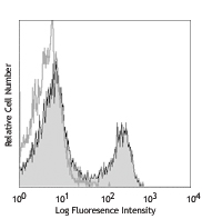
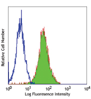
Con A-stimulated (3 days) C57BL/6 mouse splenocytes were fixed and permeabilized with 70% ethanol, then stained with Ki-67 (clone 16A8) purified antibody (filled histogram) or rat IgG2a, κ isotype control (open histogram) followed by anti-rat IgG PE. -

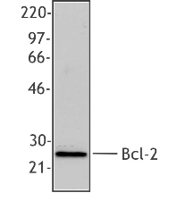
Total cell lysates (15 µg protein) from Raw264.7 and NIH3T3 were resolved by 3-8% Tris-Acetate gel electrophoresis, transferred to nitrocellulose, and probed with mouse Ki-67 antibody (clone 16A8). Proteins were visualized using a goat anti-rat IgG secondary antibody conjugated to HRP and chemiluminescence detection. Direct-Blot™ HRP anti-β-actin was used as a loading control. -

TE‐71 cells were fixed with 1% paraformaldehyde (PFA) for ten minutes, permeabilized with 0.5% Triton X‐100 for ten minutes, and blocked with 5% FBS for 30 minutes. Then the cells were intracellularly stained with 2.5 μg/mL of purified Ki‐67 (clone 16A8) in 5% FBS overnight at 4°C, followed by Alexa Fluor® 647 goat anti-rat IgG (clone Poly4054, red) for two hours. Nuclei were counterstained with DAPI (blue). The image was captured with a 40X objective. -

C57BL/6 mouse frozen intestine section was fixed with 4% paraformaldehyde (PFA) for ten minutes, permeabilized with 0.5% Triton X-100 for ten minutes, and blocked with 5% FBS plus 5% goat serum for 30 minutes at room temperature. Then the section was stained with 5 µg/mL purified Ki-67 (clone 16A8) in 5% FBS overnight at 4°C, followed by Alexa Fluor® 647 goat anti-rat IgG (clone Poly4054, red). Nuclei were counterstained with DAPI (blue). The image was captured with a 10X objective.
| Cat # | Size | Price | Quantity Check Availability | Save | ||
|---|---|---|---|---|---|---|
| 652401 | 25 µg | 89 CHF | ||||
| 652402 | 100 µg | 206 CHF | ||||
The nuclear protein Ki-67 was first identified by the monoclonal antibody Ki-67, which was generated by immunizing mice with nuclei of the L428 Hodgkin lymphoma cell line. Ki-67 protein plays an essential role in ribosomal RNA transcription and cell proliferation. Expression of Ki-67 occurs during G1, S, G2, and M phase, while in G0 phase the Ki-67 protein is not detectable. Ki-67 is strongly expressed in proliferating cells and has been reported as a prognostic marker in various tumors.
Product DetailsProduct Details
- Verified Reactivity
- Mouse
- Antibody Type
- Monoclonal
- Host Species
- Rat
- Immunogen
- E. coli expressed partial mouse Ki-67 recombinant protein, 1816-2163 aa.
- Formulation
-
This antibody is provided in phosphate-buffered solution, pH 7.2, containing 0.09% sodium azide.
Previous lots of this product may have been formulated with 0.1% or 0.05% NaN3 instead of 0.09% NaN3. For further information please contact BioLegend Technical Support or Customer Service. - Preparation
- The antibody was purified by affinity chromatography.
- Concentration
- 0.5 mg/ml
- Storage & Handling
- Upon receipt, store undiluted between 2°C and 8°C.
- Application
-
FC - Quality tested
WB, ICC, IHC-F - Verified - Recommended Usage
-
Each lot of this antibody is quality control tested by immunofluorescent staining with flow cytometric analysis. For flow cytometric staining, the suggested use of this reagent is ≤ 0.5 µg per million cells in 100 µl volume. For western blotting, suggested working dilution(s): Use 5 µl per 5 ml antibody dilution buffer for each mini-gel (0.5 µg/ml). For immunocytochemistry, a concentration range of 1.25 - 5.0 μg/mL is recommended. For immunohistochemistry on frozen tissue sections, a concentration range of 2.5 - 10.0 µg/mL is suggested. Additionally, each lot of this antibody is quality control tested by our Ki-67 staining protocol below. It is recommended that the reagent be titrated for optimal performance for each application.
-
Application References
(PubMed link indicates BioLegend citation) - Product Citations
-
- RRID
-
AB_11203533 (BioLegend Cat. No. 652401)
AB_11204254 (BioLegend Cat. No. 652402)
Antigen Details
- Structure
- 325 kD protein containing a forkhead-associated domain (FHA) and 13 tandem repeats
- Distribution
-
Nucleus and chromosome
- Function
- Required for cell cycle progression and proliferation
- Interaction
- Interacts with KIF15; binds to MKI67IP through FHA domain
- Biology Area
- Cell Biology, Cell Cycle/DNA Replication, Transcription Factors
- Molecular Family
- Nuclear Markers
- Antigen References
-
1. Starborg M, et al. 1996. J. Cell. Sci. 109:143.
2. Byeon IJ, et al. 2005. Nat. Struct. Mol. Biol. 12:987.
3. Yerushalmi R, et al. 2010. Lancet. Oncol. 11:174.
4. Beltrami AP, et al. 2001. N. Engl. J. Med. 344:1750.
5. Sachsenberg N, et al. 1998. J. Exp. Med. 187:1295.
6. Nagy Z, et al. 1997. Acta. Neuropathol. 93:294. - Gene ID
- 17345 View all products for this Gene ID
- UniProt
- View information about Ki-67 on UniProt.org

Related FAQs
Other Formats
View All Ki-67 Reagents Request Custom Conjugation| Description | Clone | Applications |
|---|---|---|
| Purified anti-mouse Ki-67 | 16A8 | FC,WB,ICC,IHC-F |
| PE anti-mouse Ki-67 | 16A8 | ICFC |
| APC anti-mouse Ki-67 | 16A8 | ICFC |
| Alexa Fluor® 647 anti-mouse Ki-67 | 16A8 | ICFC |
| FITC anti-mouse Ki-67 | 16A8 | ICFC |
| Brilliant Violet 421™ anti-mouse Ki-67 | 16A8 | ICFC |
| Brilliant Violet 605™ anti-mouse Ki-67 | 16A8 | ICFC |
| Alexa Fluor® 488 anti-mouse Ki-67 | 16A8 | ICFC |
| Alexa Fluor® 700 anti-mouse Ki-67 | 16A8 | ICFC |
| Pacific Blue™ anti-mouse Ki-67 | 16A8 | ICFC |
| PerCP/Cyanine5.5 anti-mouse Ki-67 | 16A8 | ICFC |
| PE/Cyanine7 anti-mouse Ki-67 | 16A8 | ICFC |
| PE/Dazzle™ 594 anti-mouse Ki-67 | 16A8 | ICFC |
| APC/Fire™ 750 anti-mouse Ki-67 | 16A8 | ICFC |
| Spark NIR™ 685 anti-mouse Ki-67 | 16A8 | ICFC |
Customers Also Purchased
Compare Data Across All Formats
This data display is provided for general comparisons between formats.
Your actual data may vary due to variations in samples, target cells, instruments and their settings, staining conditions, and other factors.
If you need assistance with selecting the best format contact our expert technical support team.
-
Purified anti-mouse Ki-67

Con A-stimulated (3 days) C57BL/6 mouse splenocytes were fix... 
Total cell lysates (15 µg protein) from Raw264.7 and NIH3T3 ... 
TE‐71 cells were fixed with 1% paraformaldehyde (PFA) for te... 
C57BL/6 mouse frozen intestine section was fixed with 4% par... -
PE anti-mouse Ki-67

C57BL/6 mouse splenocytes were stimulated for 3 days with Co... -
APC anti-mouse Ki-67

Con A+IL-2 stimulated (3 days) C57BL/6 mouse splenocytes wer... -
Alexa Fluor® 647 anti-mouse Ki-67

Con A-stimulated (3 days) BALB/c mouse splenocytes were fixe... -
FITC anti-mouse Ki-67

Con A-stimulated (3 days) BALB/c mouse splenocytes were fixe... -
Brilliant Violet 421™ anti-mouse Ki-67

Con A+IL-2-stimulated (3 days) C57BL/6 mouse splenocytes wer... -
Brilliant Violet 605™ anti-mouse Ki-67

Con A+IL-2-stimulated (3 days) C57BL/6 mouse splenocytes wer... -
Alexa Fluor® 488 anti-mouse Ki-67

Con A-stimulated (3 days) C57BL/6 mouse splenocytes were fix... -
Alexa Fluor® 700 anti-mouse Ki-67

Con A+IL-2 stimulated (3 days) C57BL/6 mouse splenocytes wer... -
Pacific Blue™ anti-mouse Ki-67

Con A-stimulated (three days) C57BL/6 mouse splenocytes were... -
PerCP/Cyanine5.5 anti-mouse Ki-67

Con A+IL-2-stimulated (day 3) C57BL/6 mouse splenocytes were... -
PE/Cyanine7 anti-mouse Ki-67

Con A+IL-2 stimulated (3 days) C57BL/6 mouse splenocytes wer... -
PE/Dazzle™ 594 anti-mouse Ki-67

Con A + IL-2 stimulated (three days) C57BL/6 mouse splenocyt... -
APC/Fire™ 750 anti-mouse Ki-67

Con A-stimulated (2 days) C57BL/6 mouse splenocytes were fix... -
Spark NIR™ 685 anti-mouse Ki-67

Con A+IL-2 stimulated (2 days) C57BL/6 mouse splenocytes wer...


Follow Us