 Login / Register


- Clone
- 5D3-F7 (See other available formats)
- Regulatory Status
- RUO
- Other Names
- Monocyte chemoattractant protein-1, Monocyte chemoattractant and activating factor (MCAF), JE, Small inducible cytokine A2 (SCYA2), HC-11, P6, Smooth muscle cell chemotactic factor (SMC-CF), CCL2
- Isotype
- Mouse IgG1, κ
- Ave. Rating
- Submit a Review
- Product Citations
- publications

-

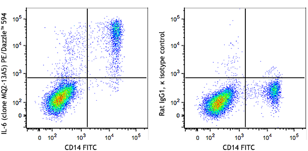
IFN-γ primed human peripheral blood monocytes were stimulated with LPS overnight (in the presence of monensin), then surface stained with CD14 APC and intracellularly stained with MCP-1 PE/Cyanine7 (left) or mouse IgG1, κ PE/Cyanine7 isotype control (right).
| Cat # | Size | Price | Quantity Check Availability | Save | ||
|---|---|---|---|---|---|---|
| 502613 | 25 tests | 147 CHF | ||||
| 502614 | 100 tests | 364 CHF | ||||
Monocyte chemotactic protein-1 (MCP-1) also known as monocyte chemotactic and activating factor (MCAF) was identified based on its ability to chemoattract monocytes. Subsequently, MCP-1 has also been found to regulate adhesion molecule expression and cytokine production in monocytes. MCP-1 is identical to the product of the JE gene, a PDGF inducible gene. MCP-1 is a member of the beta (C-C) chemokine subfamily, known as CCL2. The 5D3-F7 antibody reacts with human monocyte chemoattractant protein-1 (MCP-1).
Product DetailsProduct Details
- Verified Reactivity
- Human
- Reported Reactivity
- Cynomolgus, Rhesus
- Antibody Type
- Monoclonal
- Host Species
- Mouse
- Immunogen
- Recombinant human MCP-1
- Formulation
- Phosphate-buffered solution, pH 7.2, containing 0.09% sodium azide and BSA (origin USA)
- Preparation
- The antibody was purified by affinity chromatography and conjugated with PE/Cyanine7 under optimal conditions.
- Concentration
- Lot-specific (to obtain lot-specific concentration and expiration, please enter the lot number in our Certificate of Analysis online tool.)
- Storage & Handling
- The antibody solution should be stored undiluted between 2°C and 8°C, and protected from prolonged exposure to light. Do not freeze.
- Application
-
ICFC - Quality tested
- Recommended Usage
-
Each lot of this antibody is quality control tested by intracellular immunofluorescent staining with flow cytometric analysis. For flow cytometric staining, the suggested use of this reagent is 5 µl per million cells in 100 µl staining volume or 5 µl per 100 µl of whole blood.
- Excitation Laser
-
Blue Laser (488 nm)
Green Laser (532 nm)/Yellow-Green Laser (561 nm)
- Application Notes
-
ELISA or ELISPOT Detection1: The biotinylated 5D3-F7 antibody is useful as the detection antibody in a sandwich ELISA or ELISPOT assay, when used in conjunction with the purified 2H5 antibody (Cat. No. 505902/505906) as the capture antibody.
ELISA or ELISPOT Capture: The purified 5D3-F7 antibody is useful as the capture antibody in a sandwich ELISA or ELISPOT assay, when used in conjunction with the biotinylated 2H5 antibody (Cat. No. 505908) as the detection antibody. The LEAF™ purified antibody (Cat. No. 502607) is suggested for ELISPOT capture.
Additional reported applications (for the relevant formats) include: intracellular flow cytometry2, immunoprecipitation1,3, Western blotting1, and immunohistochemical staining1. - Additional Product Notes
-
BioLegend is in the process of converting the name PE/Cy7 to PE/Cyanine7. The dye molecule remains the same, so you should expect the same quality and performance from our PE/Cyanine7 products. Please contact Technical Service if you have any questions.
-
Application References
(PubMed link indicates BioLegend citation) -
- Peri, G., et al. 1994. J. Immunol. Meth. 174:249. (IP, IHC, WB)
- Rezaie-Majd, A., et al. 2002. Arterioscler Thromb Vasc Biol. 22:1194. (ICFC)
- Hirsch, A., et al. 1999. J. Virol. 73:404. (IP)
- Product Citations
-
- RRID
-
AB_2734490 (BioLegend Cat. No. 502613)
AB_2734491 (BioLegend Cat. No. 502614)
Antigen Details
- Structure
- CC chemokine; dimer; 13.8 kD (Mammalian)
- Bioactivity
- Chemoattractant; regulate adhesion molecule expression, cytokine production in monocytes; proliferation/activation of CC-chemokine activated killer cells
- Cell Sources
- Monocytes, vascular endothelial cells, smooth muscle, glomerular mesangial cells
- Cell Targets
- Monocytes, basophils, T cells, NK cells, immature dendritic cells
- Receptors
- CCR2; CCR5; CCR10; D6; possibly L-CCR
- Cell Type
- Embryonic Stem Cells
- Biology Area
- Cell Biology, Immunology, Neuroinflammation, Neuroscience, Stem Cells
- Molecular Family
- Cytokines/Chemokines
- Antigen References
-
1. Fitzgerald K, et al. Eds. 2001. The Cytokine FactsBook. Academic Press, San Diego.
2. Bischoff S, et al. 1992. J. Exp. Med. 175:1271.
3. Charo I, et al. 1994. P. Natl. Acad. Sci. USA 91:2752.
4. Jiang Y, et al. 1992. J. Immunol. 148:2423. - Regulation
- Upregulated by IL-1, TNF-α, PDGF, TGF-β
- Gene ID
- 6347 View all products for this Gene ID
- UniProt
- View information about CCL2 on UniProt.org
Related Pages & Pathways
Pathways


Related FAQs
Other Formats
View All CCL2 Reagents Request Custom Conjugation| Description | Clone | Applications |
|---|---|---|
| PE anti-human MCP-1 | 5D3-F7 | ICFC |
| Purified anti-human MCP-1 | 5D3-F7 | ELISA Capture,WB,ELISA,ELISPOT Detection,ICFC,IHC,IP |
| Biotin anti-human MCP-1 | 5D3-F7 | ELISA Detection,ELISPOT Detection,ICFC |
| APC anti-human MCP-1 | 5D3-F7 | ICFC |
| PE/Cyanine7 anti-human MCP-1 | 5D3-F7 | ICFC |
Customers Also Purchased

Compare Data Across All Formats
This data display is provided for general comparisons between formats.
Your actual data may vary due to variations in samples, target cells, instruments and their settings, staining conditions, and other factors.
If you need assistance with selecting the best format contact our expert technical support team.
-
PE anti-human MCP-1

IFN-γ primed human peripheral blood monocytes were stimulate... -
Purified anti-human MCP-1

Immunoprecipitation/Western blot analysis of capturing abili... 
Western blot analysis of recombinant human MCP-1 (lane 1), a... 
-
Biotin anti-human MCP-1

-
APC anti-human MCP-1

IFN-γ primed human peripheral blood monocytes were stimulate... -
PE/Cyanine7 anti-human MCP-1

IFN-γ primed human peripheral blood monocytes were sti...


Follow Us