 Login / Register


- Clone
- RMV-7 (See other available formats)
- Regulatory Status
- RUO
- Other Names
- αV integrin, Vitronectin Receptor, Integrin αV chain, ITGAV
- Isotype
- Rat IgG1, κ
- Ave. Rating
- Submit a Review
- Product Citations
- publications

-

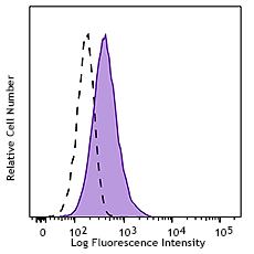
C57BL/6 mouse bone marrow cells were stained with CD51 (clone RMV-7) PE (filled histogram) or Rat IgG1, κ PE isotype control (open histogram). Data shown was gated on myeloid cell population. -

Con A stimulated C57BL/6 mouse slenocytes were stained with CD3 APC and CD51 (clone RMV-7) PE (above) or Rat IgG1, κ PE isotype control (below). -

| Cat # | Size | Price | Quantity Check Availability | Save | ||
|---|---|---|---|---|---|---|
| 104105 | 50 µg | 98 CHF | ||||
| 104106 | 200 µg | 304 CHF | ||||
CD51 is a 140 kD protein, also known as αV integrin, vitronectin receptor, and integrin αV. It is a member of the integrin family, expressed on activated T cells, polymorphonuclear granulocytes, platelets, blastocysts, and osteoclasts. CD51 forms heterodimers by association with integrins β1, β3, β5 or β6; these complexes then act as receptors for multiple extracellular matrix proteins (ECM). The αv integrin heterodimers have varied functions in development, stimulation/activation and homeostasis. The primary ligands for CD51 complexes are fibronectin, fibrinogen, vitronectin, thrombspondin, von Willebrand factor, and CD31. The RMV-7 antibody has been reported to block binding of CD51 to vitronectin, fibronectin, and CD31 in some cell types, as well as blocking LAK cell cytotoxicity.
Product DetailsProduct Details
- Verified Reactivity
- Mouse
- Antibody Type
- Monoclonal
- Host Species
- Rat
- Immunogen
- BALB/c mouse IL-2-activated killer (LAK) cells
- Formulation
- Phosphate-buffered solution, pH 7.2, containing 0.09% sodium azide.
- Preparation
- The antibody was purified by affinity chromatography, and conjugated with PE under optimal conditions.
- Concentration
- 0.2 mg/ml
- Storage & Handling
- The antibody solution should be stored undiluted between 2°C and 8°C, and protected from prolonged exposure to light. Do not freeze.
- Application
-
FC - Quality tested
- Recommended Usage
-
Each lot of this antibody is quality control tested by immunofluorescent staining with flow cytometric analysis. For flow cytometric staining, the suggested use of this reagent is ≤1.0 µg per million cells in 100 µl volume. It is recommended that the reagent be titrated for optimal performance for each application.
- Excitation Laser
-
Blue Laser (488 nm)
Green Laser (532 nm)/Yellow-Green Laser (561 nm)
- Application Notes
-
Additional reported applications (for the relevant formats) include: immunoprecipitation1, and blocking1-3 of CD51 adhesion and LAK cell cytotoxicity. The Ultra-LEAF™ purified antibody (Endotoxin <0.01 EU/µg, Azide-Free, 0.2 µm filtered) is recommended for functional assays (Cat. Nos. 104109 & 104110).
-
Application References
(PubMed link indicates BioLegend citation) -
- Takahashi K, et al. 1990. J. Immunol. 145:4371. (IP Block)
- Narumiya S, et al. 1994. Int. Immunol. 6:139. (Block)
- Piali L, et al. 1995. J. Cell Biol. 130:451. (Block)
- Delpino MV, et al. 2012. J. Leukoc Biol 91:285. PubMed.
- Product Citations
-
- RRID
-
AB_313074 (BioLegend Cat. No. 104105)
AB_2129493 (BioLegend Cat. No. 104106)
Antigen Details
- Structure
- Integrin family, associates with integrins β1, β3, β5 or β6, 140 kD
- Distribution
-
Activated T cells, polymorphonuclear granulocytes, blastocysts, osteoclasts
- Function
- Adhesion
- Ligand/Receptor
- Fibronectin, fibrinogen, vitronectin, thrombspondin, von Willebrand factor, and CD31
- Cell Type
- Granulocytes, Mesenchymal Stem Cells, Osteoclasts, T cells
- Biology Area
- Angiogenesis, Cell Adhesion, Cell Biology, Costimulatory Molecules, Immunology, Innate Immunity, Stem Cells
- Molecular Family
- Adhesion Molecules, CD Molecules
- Antigen References
-
1. Barclay AN, et al. 1997. The Leukocyte Antigen FactsBook Academic Press.
2. Maxfield SR, et al. 1989. J. Exp. Med. 169:2173.
3. Piali L, et al. 1995. J. Cell Biol. 130:451. - Gene ID
- 16410 View all products for this Gene ID
- UniProt
- View information about CD51 on UniProt.org


Related FAQs
- What type of PE do you use in your conjugates?
- We use R-PE in our conjugates.
Other Formats
View All CD51 Reagents Request Custom Conjugation| Description | Clone | Applications |
|---|---|---|
| Biotin anti-mouse CD51 | RMV-7 | FC |
| PE anti-mouse CD51 | RMV-7 | FC |
| Purified anti-mouse CD51 | RMV-7 | FC,IP,Block |
| TotalSeq™-A1008 anti-mouse CD51 | RMV-7 | PG |
| Ultra-LEAF™ Purified anti-mouse CD51 | RMV-7 | FC,IP,Block |
| TotalSeq™-C1008 anti-mouse CD51 | RMV-7 | PG |
| TotalSeq™-B1008 anti-mouse CD51 | RMV-7 | PG |
Customers Also Purchased

Compare Data Across All Formats
This data display is provided for general comparisons between formats.
Your actual data may vary due to variations in samples, target cells, instruments and their settings, staining conditions, and other factors.
If you need assistance with selecting the best format contact our expert technical support team.
-
Biotin anti-mouse CD51

C57BL/6 bone marrow cells stained with RMV-7 PE -
PE anti-mouse CD51

C57BL/6 mouse bone marrow cells were stained with CD51 (clon... 

Con A stimulated C57BL/6 mouse slenocytes were stained with ... -
Purified anti-mouse CD51

C57BL/6 mouse bone marrow granulocytes stained with CD51 (cl... -
TotalSeq™-A1008 anti-mouse CD51
-
Ultra-LEAF™ Purified anti-mouse CD51
C57BL/6 mouse bone marrow granulocytes stained with CD51 (cl... -
TotalSeq™-C1008 anti-mouse CD51
-
TotalSeq™-B1008 anti-mouse CD51


Follow Us