 Login / Register


- Clone
- 53-6.7 (See other available formats)
- Regulatory Status
- RUO
- Other Names
- T8, Lyt2, Ly-2
- Isotype
- Rat IgG2a, κ
- Ave. Rating
- Submit a Review
- Product Citations
- publications

-

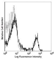
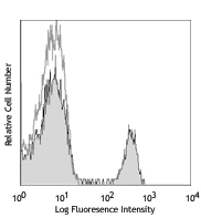
C57BL/6 mouse splenocytes were stained with CD8 (clone 53-6.7) Alexa Fluor® 647 (filled histogram) or rat IgG2a, κ Alexa Fluor® 647 isotype control (open histogram). -

C57BL/6 mouse frozen lymph node section was fixed with 4% paraformaldehyde (PFA) for 10 minutes at room temperature and blocked with 5% FBS plus 5% rat serum for 1 hour at room temperature. Then the section was stained with 5 µg/ml of B220 (clone RA3-6B2) Alexa Fluor® 594 (blue), 5 µg/ml of CD8 (clone 53-6.7) Alexa Fluor® 647 (red), and 5 µg/ml of CD4 (clone GK1.5) Alexa Fluor® 488 (green) overnight at 4ËšC. The image was captured by 10X objective. -

Paraformaldehyde-fixed (1%), 500 μm-thick mouse spleen section was processed according to the Ce3DTM Tissue Clearing Kit protocol (cat. no. 427701). The section was costained with anti-mouse CD68 Antibody (clone FA-11) Alexa Fluor® 488 at 5 µg/mL (green), and anti-mouse CD8a Antibody (clone 53-6.7) Alexa Fluor® 647 at 5 µg/mL (magenta) and counterstained with DAPI (blue). The section was then optically cleared and mounted in a sample chamber. The image was captured with a 10X objective using Zeiss 780 confocal microscope and processed by Imaris image analysis software.
Watch the video. -

Confocal image of C57BL/6 mouse lung sample acquired using the IBEX method of highly multiplexed antibody-based imaging: Ly-6G (yellow) in Cycle 2, CD31 (blue) in Cycle 3, and CD8 (red) in Cycle 4. Tissues were prepared using ~1% (vol/vol) formaldehyde and a detergent. Following fixation, samples are immersed in 30% (wt/vol) sucrose for cryoprotection. Images are courtesy of Drs. Andrea J. Radtke and Ronald N. Germain of the Center for Advanced Tissue Imaging (CAT-I) in the National Institute of Allergy and Infectious Diseases (NIAID, NIH).
| Cat # | Size | Price | Quantity Check Availability | Save | ||
|---|---|---|---|---|---|---|
| 100727 | 25 µg | 101 CHF | ||||
| 100724 | 100 µg | 216 CHF | ||||
CD8, also known as Lyt-2, Ly-2, or T8, consists of disulfide-linked α and β chains that form the α(CD8a)/β(CD8b) heterodimer and α/α homodimer. CD8a is a 34 kD protein that belongs to the immunoglobulin family. The CD8 α/β heterodimer is expressed on the surface of most thymocytes and a subset of mature TCR α/β T cells. CD8 expression on mature T cells is non-overlapping with CD4. The CD8 α/α homodimer is expressed on a subset of γ/δ TCR-bearing T cells, NK cells, intestinal intraepithelial lymphocytes, and lymphoid dendritic cells. CD8 is an antigen co-receptor on T cells that interacts with MHC class I on antigen-presenting cells or epithelial cells. CD8 promotes T cell activation through its association with the TCR complex and protein tyrosine kinase lck.
Product DetailsProduct Details
- Verified Reactivity
- Mouse
- Antibody Type
- Monoclonal
- Host Species
- Rat
- Immunogen
- Mouse thymus or spleen
- Formulation
- Phosphate-buffered solution, pH 7.2, containing 0.09% sodium azide.
- Preparation
- The antibody was purified by affinity chromatography and conjugated with Alexa Fluor® 647 under optimal conditions.
- Concentration
- 0.5 mg/mL
- Storage & Handling
- The antibody solution should be stored undiluted between 2°C and 8°C, and protected from prolonged exposure to light. Do not freeze.
- Application
-
FC - Quality tested
IHC-F, 3D IHC - Verified
SB - Community verified
SB - Reported in the literature, not verified in house - Recommended Usage
-
Each lot of this antibody is quality control tested by immunofluorescent staining with flow cytometric analysis. For flow cytometric staining, the suggested use of this reagent is ≤ 0.25 µg per million cells in 100 µL volume. For immunohistochemistry on frozen tissue sections, a concentration range of 2.5 - 5.0 μg/mL is suggested. For 3D immunohistochemistry on formalin-fixed tissues, a concentration of 5.0 µg/mL is suggested. It is recommended that the reagent be titrated for optimal performance for each application.
* Alexa Fluor® 647 has a maximum emission of 668 nm when it is excited at 633nm / 635nm.
Alexa Fluor® and Pacific Blue™ are trademarks of Life Technologies Corporation.
View full statement regarding label licenses - Excitation Laser
-
Red Laser (633 nm)
- Application Notes
-
Clone 53-6.7 antibody competes with clone 5H10-1 antibody for binding to thymocytes3. The 53-6.7 antibody has been reported to block antigen presentation via MHC class I and inhibit T cell responses to IL-2. This antibody has also been used for depletion of CD8a+ cells. Additional reported applications (for the relevant formats) include: immunoprecipitation1,3, in vivo and in vitro cell depletion2,10,15, inhibition of CD8 T cell proliferation3, blocking of cytotoxicity3,4, immunohistochemical staining5,6 of acetone-fixed frozen sections and zinc-fixed paraffin-embedded sections, and spatial biology (IBEX)29,30. Clone 53-6.7 is not recommended for immunohistochemistry of formalin-fixed paraffin sections. The Ultra-LEAF™ purified antibody (Endotoxin < 0.01 EU/µg, Azide-Free, 0.2 µm filtered) is recommended for functional assays or in vivo studies (Cat No. 100746).
- Additional Product Notes
-
For use in spatial biology, this antibody has been demonstrated for use in immunohistochemistry using IBEX (Reported in the literature, not verified in house) and the NanoString GeoMx® Digital Spatial Profiler.
IBEX: Iterative Bleaching Extended multi-pleXity (IBEX) is a fluorescent imaging technique capable of highly-multiplexed spatial analysis. The method relies on cyclical bleaching of panels of fluorescent antibodies in order to image and analyze many markers over multiple cycles of staining, imaging, and, bleaching. It is a community-developed open-access method developed by the Center for Advanced Tissue Imaging (CAT-I) in the National Institute of Allergy and Infectious Diseases (NIAID, NIH).
NanoString GeoMx®: This product has been verified for IHC-F (Immunohistochemistry - frozen tissue sections) on the NanoString GeoMx® Digital Spatial Profiler. The GeoMx® enables researchers to perform spatial analysis of protein and RNA targets in FFPE and fresh frozen human and mouse samples. For more information about our spatial biology products and the GeoMx® platform, please visit our spatial biology page. -
Application References
(PubMed link indicates BioLegend citation) -
- Ledbetter JA, et al. 1979. Immunol. Rev. 47:63. (IHC, IP)
- Hathcock KS. 1991. Current Protocols in Immunology. 3.4.1. (Deplete)
- Takahashi K, et al. 1992. P. Natl. Acad. Sci. USA 89:5557. (Block, IP)
- Ledbetter JA, et al. 1981. J. Exp. Med. 153:1503. (Block)
- Hata H, et al. 2004. J. Clin. Invest. 114:582. (IHC)
- Fan WY, et al. 2001. Exp. Biol. Med. 226:1045. (IHC)
- Shih FF, et al. 2006. J. Immunol. 176:3438. (FC)
- Kamimura D, et al. 2006. J. Immunol. 177:306.
- Bouwer HGA, et al. 2006. P. Natl. Acad. Sci. USA 103:5102. (FC, Deplete)
- Kao C, et al. 2005. Int. Immunol. 17:1607. PubMed
- Ko SY, et al. 2005. J. Immunol. 175:3309. (FC) PubMed
- Rasmussen JW, et al. 2006. Infect. Immun. 74:6590. PubMed
- Lee CH, et al. 2009. Clin. Cancer Res. PubMed
- Geiben-Lynn R, et al. 2008. Blood 112:4585. (Deplete) PubMed
- Kingeter LM, et al. 2008. J. Immunol. 181:6244. PubMed
- Guo Y, et al. 2008. Blood 112:480. PubMed
- Andrews DM, et al. 2008. J. Virol. 82:4931. PubMed
- Britschqui MR, et al. 2008. J. Immunol. 181:7681. PubMed
- Kenna TJ, et al. 2008. Blood 111:2091. PubMed
- Jordan JM, et al. 2008. Infect. Immun. 76:3717. PubMed
- Todd DJ, et al. 2009. J. Exp. Med. 206:2151. PubMed
- Bankoti J, et al. 2010. Toxicol. Sci. 115:422. (FC) PubMed
- Medyouf H, et al. 2010. Blood 115:1175. PubMed
- Riedl P, et al. 2009. J. Immunol. 183:370. PubMed
- Apte SH, et al. 2010. J. Immunol. 185:998. PubMed
- Bankoti J, et al. 2010. Toxicol. Sci. 115:422. (FC) PubMed
- del Rio ML, et al. 2011. Transpl. Int. 24:501. (FC) PubMed
- Cui L, et al. 2015. J Control Release. 206:220. PubMed
- Radtke AJ, et al. 2020. Proc Natl Acad Sci U S A. 117:33455-65. (SB) PubMed
- Radtke AJ, et al. 2022. Nat Protoc. 17:378-401. (SB) PubMed
- Product Citations
-
- RRID
-
AB_493424 (BioLegend Cat. No. 100727)
AB_389326 (BioLegend Cat. No. 100724)
Antigen Details
- Structure
- Ig superfamily, CD8α chain, 34 kD
- Distribution
-
Most thymocytes, T cell subset, some NK cells, lymphoid dendritic cells
- Function
- Co-receptor for TCR
- Ligand/Receptor
- MHC class I molecule
- Antigen References
-
1. Barclay A, et al. 1997. The Leukocyte Antigen FactsBook Academic Press.
2. Zamoyska R. 1994. Immunity 1:243.
3. Ellmeier W, et al. 1999. Annu. Rev. Immunol. 17:523. - Gene ID
- 12525 View all products for this Gene ID
- UniProt
- View information about CD8alpha on UniProt.org


Related FAQs
- If an antibody clone has been previously successfully used in IBEX in one fluorescent format, will other antibody formats work as well?
-
It’s likely that other fluorophore conjugates to the same antibody clone will also be compatible with IBEX using the same sample fixation procedure. Ultimately a directly conjugated antibody’s utility in fluorescent imaging and IBEX may be specific to the sample and microscope being used in the experiment. Some antibody clone conjugates may perform better than others due to performance differences in non-specific binding, fluorophore brightness, and other biochemical properties unique to that conjugate.
- Will antibodies my lab is already using for fluorescent or chromogenic IHC work in IBEX?
-
Fundamentally, IBEX as a technique that works much in the same way as single antibody panels or single marker IF/IHC. If you’re already successfully using an antibody clone on a sample of interest, it is likely that clone will have utility in IBEX. It is expected some optimization and testing of different antibody fluorophore conjugates will be required to find a suitable format; however, legacy microscopy techniques like chromogenic IHC on fixed or frozen tissue is an excellent place to start looking for useful antibodies.
- Are other fluorophores compatible with IBEX?
-
Over 18 fluorescent formats have been screened for use in IBEX, however, it is likely that other fluorophores are able to be rapidly bleached in IBEX. If a fluorophore format is already suitable for your imaging platform it can be tested for compatibility in IBEX.
- The same antibody works in one tissue type but not another. What is happening?
-
Differences in tissue properties may impact both the ability of an antibody to bind its target specifically and impact the ability of a specific fluorophore conjugate to overcome the background fluorescent signal in a given tissue. Secondary stains, as well as testing multiple fluorescent conjugates of the same clone, may help to troubleshoot challenging targets or tissues. Using a reference control tissue may also give confidence in the specificity of your staining.
- How can I be sure the staining I’m seeing in my tissue is real?
-
In general, best practices for validating an antibody in traditional chromogenic or fluorescent IHC are applicable to IBEX. Please reference the Nature Methods review on antibody based multiplexed imaging for resources on validating antibodies for IBEX.
Other Formats
View All CD8a Reagents Request Custom ConjugationCustomers Also Purchased
Compare Data Across All Formats
This data display is provided for general comparisons between formats.
Your actual data may vary due to variations in samples, target cells, instruments and their settings, staining conditions, and other factors.
If you need assistance with selecting the best format contact our expert technical support team.
-
APC anti-mouse CD8a

C57BL/6 mouse splenocytes were stained with CD8 (clone 53-6.... -
Biotin anti-mouse CD8a

C57BL/6 mouse splenocytes were stained with biotinylated CD8... -
FITC anti-mouse CD8a

C57BL/6 mouse splenocytes were stained with CD8 (clone 53-6.... -
PE anti-mouse CD8a

C57BL/6 mouse splenocytes were stained with CD8 (clone 53-6.... 
C57BL/6 mouse splenocytes were stained with CD8a (clone 53-6... -
PE/Cyanine5 anti-mouse CD8a

C57BL/6 splenocytes were stained with CD8 (clone 53-6.7) PE/... -
Purified anti-mouse CD8a

C57BL/6 mouse splenocytes were stained with purified CD8 (cl... 
C57BL/6 frozen mouse spleen section was fixed with 4% parafo... 
Fresh, frozen mouse spleen was stained with purified CD8a cl... -
PE/Cyanine7 anti-mouse CD8a

C57BL/6 splenocytes were stained with CD8 (clone 53-6.7) PE/... -
APC/Cyanine7 anti-mouse CD8a

C57BL/6 mouse splenocytes were stained with CD3 FITC and CD8... -
Alexa Fluor® 488 anti-mouse CD8a

C57BL/6 mouse splenocytes were stained with CD8 (clone 53-6.... 
Paraformaldehyde-fixed (1%), 500 µm-thick mouse thymus secti... -
Alexa Fluor® 647 anti-mouse CD8a

C57BL/6 mouse splenocytes were stained with CD8 (clone 53-6.... 
C57BL/6 mouse frozen lymph node section was fixed with 4% pa... 
Paraformaldehyde-fixed (1%), 500 μm-thick mouse spleen secti... 
Confocal image of C57BL/6 mouse lung sample acquired using t... -
Pacific Blue™ anti-mouse CD8a

C57BL/6 mouse splenocytes were stained with CD8 (clone 53-6.... -
Alexa Fluor® 700 anti-mouse CD8a

C57BL/6 mouse splenocytes stained with CD8 (clone 53-6.7) Al... -
PerCP/Cyanine5.5 anti-mouse CD8a

C57BL/6 mouse splenocytes were stained with CD3ε FIT... -
PerCP anti-mouse CD8a

C57BL/6 thymocytes were stained with CD8 (clone 53-6.7) PerC... -
Brilliant Violet 421™ anti-mouse CD8a

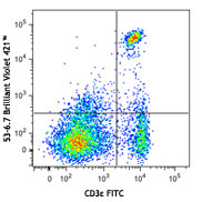
C57BL/6 mouse splenocytes were stained with CD3ε FITC and CD... 
BL6 mouse lymph nodes, fixed O/N in PLP, blocked with 10% ra... 
Confocal image of C57BL/6 mouse spleen sample acquired using... 
Confocal image of C57BL/6 mouse thymus sample acquired using... 
Confocal image of C57BL/6 mouse small intestine sample acqui... 
Confocal image of C57BL/6 mouse liver sample acquired using ... -
Brilliant Violet 570™ anti-mouse CD8a

C57BL/6 mouse splenocytes were stained with CD3 FITC and CD8... 
-
Brilliant Violet 650™ anti-mouse CD8a

C57BL/6 mouse splenocytes were stained with CD3 PE and CD8a ... -
Brilliant Violet 605™ anti-mouse CD8a

C57BL/6 mouse splenocytes were stained with CD3 FITC and CD8... -
Ultra-LEAF™ Purified anti-mouse CD8a

C57BL/6 mouse splenocytes were stained with LEAF™ purified C... -
Brilliant Violet 711™ anti-mouse CD8a

C57BL/6 mouse splenocytes were stained with CD3 PE and CD8a ... -
Brilliant Violet 785™ anti-mouse CD8a

C57BL/6 mouse splenocytes were stained with CD3 PE and CD8a ... -
Brilliant Violet 510™ anti-mouse CD8a

C57BL/6 mouse splenocytes were stained with CD3 APC and CD8a... -
Purified anti-mouse CD8a (Maxpar® Ready)

C57BL/6 mouse splenocytes stained with 147Sm-anti-CD45 (30-F... -
Alexa Fluor® 594 anti-mouse CD8a

C57BL/6 mouse frozen lymph node section was fixed with 4% pa... 
C57BL/6 mouse frozen lymph node section was fixed with 4% pa... 
Paraformaldehyde-fixed (1%), 500 µm-thick mouse thymus secti... -
PE/Dazzle™ 594 anti-mouse CD8a

C57BL/6 mouse splenocytes were stained with CD3ε FITC and C... 
-
APC/Fire™ 750 anti-mouse CD8a

C57BL/6 thymocytes were stained with CD4 PE and CD8a (clone ... 
-
GoInVivo™ Purified anti-mouse CD8a
-
TotalSeq™-A0002 anti-mouse CD8a
-
Spark Blue™ 550 anti-mouse CD8a

C57BL/6 mouse splenocytes were stained with CD8 (clone 53-6.... -
Spark NIR™ 685 anti-mouse CD8a

C57BL/6 mouse splenocytes were stained with CD8a (clone 53-6... -
TotalSeq™-C0002 anti-mouse CD8a
-
TotalSeq™-B0002 anti-mouse CD8a
-
Spark YG™ 570 anti-mouse CD8a

C57BL/6 mouse frozen spleen section was fixed with 4% parafo... -
PE/Fire™ 640 anti-mouse CD8a

C57BL/6 mouse splenocytes were stained with CD3 Alexa Fluor®... -
PE/Fire™ 700 anti-mouse CD8a

C57BL/6 mouse splenocytes were stained with anti-mouse CD3 A... -
Spark Blue™ 574 anti-mouse CD8a Antibody

C57BL/6 splenocytes were stained with anti-mouse CD3e APC an... -
Spark Violet™ 423 anti-mouse CD8a Antibody

C57BL/6 splenocytes were stained with anti-mouse CD3 PE and ... -
Spark UV™ 387 anti-mouse CD8a

C57BL/6 mouse splenocytes were stained with anti-mouse CD3ε ... -
Spark Blue™ 515 anti-mouse CD8a

C57BL/6 mouse splenocytes were stained with anti-mouse CD3ε ... -
APC/Fire™ 810 anti-mouse CD8a

C57BL/6 mouse splenocytes were stained with anti-mouse CD3&e... -
Spark Red™ 718 anti-mouse CD8a (Flexi-Fluor™)
-
Spark PLUS UV™ 395 anti-mouse CD8a

C57BL/6 mouse splenocytes were stained with anti-mouse CD3ε ...


Follow Us