 Login / Register


- Clone
- Ki-67 (See other available formats)
- Regulatory Status
- RUO
- Other Names
- Antigen Ki-67
- Isotype
- Mouse IgG1, κ
- Ave. Rating
- Submit a Review
- Product Citations
- publications

-

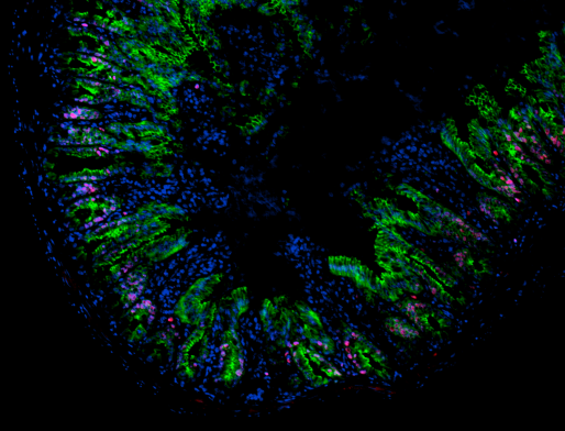
HeLa cells were fixed with 1% paraformaldehyde (PFA) for 10 minutes, permeabilized with 0.5% Triton X-100 for 10 minutes, and blocked with 5% FBS for 30 minutes. Then the cells were intracellularly stained with 5 µg/ml anti-human Ki-67 (clone Ki-67) Alexa Fluor® 594 (red) in blocking buffer overnight at 4°C and followed by Alexa Fluor® 488 Phalloidin (green) staining for 20 minutes at 4°C. Nuclei were counterstained with DAPI (blue). The image was captured with a 20X objective.
| Cat # | Size | Price | Quantity Check Availability | Save | ||
|---|---|---|---|---|---|---|
| 350528 | 100 µg | 323 CHF | ||||
Antigen Ki-67 is a nuclear protein expressed as two isoforms with molecular weights of 395 and 345 kD. Both isoforms contain one forkhead-associated domain and 16 concatenated "Ki-67 repeats," each containing the epitope recognized by the mAb Ki-67. The antigen Ki-67 interacts with Hklp2, hNIFK, and chromobox protein homolog 1, 3, and 5. Ki-67 is required for cell proliferation and its expression is restricted to the phases G1, S, G2, and M of the cell cycle. This characteristic makes Ki-67 an excellent marker for proliferating cells and is commonly used as one of the prognostic factors in cancer studies. Ki-67 has also been used to study myocyte proliferation after myocardial infarction as well as lymphocyte proliferation during infection, and has been used in neurons of patients with different neuropathologies.
Product DetailsProduct Details
- Verified Reactivity
- Human
- Reported Reactivity
- Cow
- Antibody Type
- Monoclonal
- Host Species
- Mouse
- Immunogen
- Nuclei of the Hodgkin lymphoma cell line L428
- Formulation
- Phosphate-buffered solution, pH 7.2, containing 0.09% sodium azide.
- Preparation
- The antibody was purified by affinity chromatography and conjugated with Alexa Fluor® 594 under optimal conditions.
- Concentration
- 0.5 mg/ml
- Storage & Handling
- The antibody solution should be stored undiluted between 2°C and 8°C, and protected from prolonged exposure to light. Do not freeze.
- Application
-
ICC - Quality tested
- Recommended Usage
-
Each lot of this antibody is quality control tested by immunocytochemistry. For immunocytochemistry, a concentration range of 1.0 - 5.0 μg/ml is recommended. It is recommended that the reagent be titrated for optimal performance for each application.
* Alexa Fluor® 594 has an excitation maximum of 590 nm, and a maximum emission of 617 nm.
Alexa Fluor® and Pacific Blue™ are trademarks of Life Technologies Corporation.
View full statement regarding label licenses - Application Notes
-
Additional reported applications (for the relevant formats) include: immunohistochemical staining of frozen tissue sections1, Western blotting3, and immunofluorescence microscopy4.
Ki-67 Staining Protocol:
1. Prepare 70% ethanol and chill at -20°C.
2. Prepare target cells of interest and wash 2X with PBS by centrifuge at 350xg for 5 minutes.
3. Discard supernatant and loosen the cell pellet by vortexing.
4. Add 3 ml cold 70% ethanol drop by drop to the cell pellet while vortexing.
5. Continue vortexing for 30 seconds and then incubate at -20°C for 1 hour.
6. Wash 3X with BioLegend Cell Staining Buffer and then resuspend the cells at the concentration of 0.5-10 x 106/ml.
7. Mix 100 µl cell suspension with proper fluorochrome-conjugated Ki-67 antibody and incubate at room temperature in the dark for 30 minutes.
8. Wash 2X with BioLegend Cell Staining Buffer and then resuspend in 0.5 ml cell staining buffer for flow cytometric analysis. -
Application References
(PubMed link indicates BioLegend citation) -
- Gerdes J, et al. 1983. Int. J. Cancer 31:13. (IHC)
- Gerdes J, et al. 1984. J. Immunol. 133:1710. (ICFC)
- Schluter C, et al. 1993 J. Cell Biol. 123:513. (IHC, WB)
- Bading H, et al. 1989 Exp. Cell. Res. 185:50. (IF)
- Guha P, et al. 2013. PNAS. 110:5052. PubMed
- Product Citations
-
- RRID
-
AB_2563504 (BioLegend Cat. No. 350528)
Antigen Details
- Structure
- Two isoforms with molecular weights of 395 and 345 kD, one forkhead-associated domain, 16 concatenated Ki-67 repeats, located in nucleus
- Distribution
-
Expressed in the phases G1, S, G2, and M of the cell cycle
- Function
- Required for cell proliferation
- Interaction
- Chromobox protein homolog 1, 3 and 5, Hklp2, and hNIFK
- Biology Area
- Cell Biology, Cell Cycle/DNA Replication, DNA Repair/Replication
- Molecular Family
- Nuclear Markers
- Antigen References
-
1. Byeon IJ, et al. 2005. Nat. Struct. Mol. Biol. 12:987.
2. Yerushalmi R, et al. 2010. Lancet. Oncol. 11:174.
3. Beltrami AP, et al. 2001. N. Engl. J. Med. 344:1750.
4. Sachsenberg N, et al. 1998. J. Exp. Med. 187:1295.
5. Nagy Z, et al. 1997. Acta. Neuropathol. 93:294. - Gene ID
- 4288 View all products for this Gene ID
- UniProt
- View information about Ki-67 on UniProt.org


Related FAQs
Other Formats
View All Ki-67 Reagents Request Custom Conjugation| Description | Clone | Applications |
|---|---|---|
| Brilliant Violet 510™ anti-human Ki-67 | Ki-67 | ICFC,ICC |
| Purified anti-human Ki-67 | Ki-67 | ICFC,CyTOF®,ICC,WB,IHC-F |
| PE anti-human Ki-67 | Ki-67 | ICFC |
| Brilliant Violet 421™ anti-human Ki-67 | Ki-67 | ICFC,ICC |
| Alexa Fluor® 488 anti-human Ki-67 | Ki-67 | ICFC,ICC |
| Alexa Fluor® 647 anti-human Ki-67 | Ki-67 | ICFC,ICC |
| Pacific Blue™ anti-human Ki-67 | Ki-67 | ICFC |
| APC anti-human Ki-67 | Ki-67 | ICFC |
| Brilliant Violet 711™ anti-human Ki-67 | Ki-67 | ICFC |
| PerCP/Cyanine5.5 anti-human Ki-67 | Ki-67 | ICFC |
| Brilliant Violet 605™ anti-human Ki-67 | Ki-67 | ICFC |
| PE/Cyanine7 anti-human Ki-67 | Ki-67 | ICFC |
| Purified anti-human Ki-67 (Maxpar® Ready) | Ki-67 | ICFC,CyTOF® |
| Alexa Fluor® 594 anti-human Ki-67 | Ki-67 | ICC |
| Alexa Fluor® 700 anti-human Ki-67 | Ki-67 | ICFC |
| PE/Dazzle™ 594 anti-human Ki-67 | Ki-67 | ICFC |
| Brilliant Violet 750™ anti-human Ki-67 | Ki-67 | ICFC |
Customers Also Purchased
Compare Data Across All Formats
This data display is provided for general comparisons between formats.
Your actual data may vary due to variations in samples, target cells, instruments and their settings, staining conditions, and other factors.
If you need assistance with selecting the best format contact our expert technical support team.
-
Brilliant Violet 510™ anti-human Ki-67

PHA-stimulated (3 days) human peripheral blood lymphocytes w... 
HeLa cells were fixed with 1% paraformaldehyde (PFA) for 10 ... -
Purified anti-human Ki-67

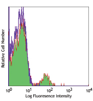
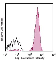
Resting (dashed line) or PHA-activated human peripheral bloo... 
HeLa cells were fixed with 1% paraformaldehyde (PFA) for 10 ... 
Total lysates (15 µg protein) from 293T control (Ctrl) and K... -
PE anti-human Ki-67

Human T leukemia cell line, Jurkat, fixed and permeabilized ...
Resting (dashed line) or PHA-activated human peripheral bloo... -
Brilliant Violet 421™ anti-human Ki-67

3-day PHA-stimulated (top) or non-stimulated (bottom) human ... 

HeLa cells were fixed with 1% paraformaldehyde (PFA) for 10 ... -
Alexa Fluor® 488 anti-human Ki-67

PHA-activated human peripheral blood lymphocytes (3 days) we... 
3-day PHA-stimulated human peripheral blood lymphocytes were... 
HeLa cells were fixed with 1% paraformaldehyde (PFA) for 10 ... -
Alexa Fluor® 647 anti-human Ki-67

PHA-stimulated (3 days) human peripheral blood lymphocytes w... 
HeLa cells were fixed with 1% paraformaldehyde (PFA) for 10 ... -
Pacific Blue™ anti-human Ki-67

PHA-stimulated (3 days) human peripheral blood lymphocytes w... -
APC anti-human Ki-67

PHA-activated human peripheral blood lymphocytes (3 days) we... 
3-day PHA-stimulated human peripheral blood lymphocytes wer... -
Brilliant Violet 711™ anti-human Ki-67

3-day PHA-stimulated (top) or non-stimulated (bottom) human ... 
-
PerCP/Cyanine5.5 anti-human Ki-67

PHA-activated human peripheral blood lymphocytes (1 day) wer... -
Brilliant Violet 605™ anti-human Ki-67

PHA-activated human peripheral blood lymphocytes (3 days) we... -
PE/Cyanine7 anti-human Ki-67

PHA-activated human peripheral blood lymphocytes (3 days) we... -
Purified anti-human Ki-67 (Maxpar® Ready)

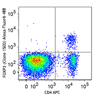
Human PBMCs were incubated for 3 days in media alone (left) ... -
Alexa Fluor® 594 anti-human Ki-67

HeLa cells were fixed with 1% paraformaldehyde (PFA) for 10 ... -
Alexa Fluor® 700 anti-human Ki-67

PHA-activated human peripheral blood lymphocytes (3 days) we... -
PE/Dazzle™ 594 anti-human Ki-67

PHA-stimulated human peripheral blood lymphocytes (three day... -
Brilliant Violet 750™ anti-human Ki-67

PHA-activated (3 days) human peripheral blood lymphocytes we...


Follow Us