 Login/Register


- Clone
- ICRF44 (See other available formats)
- Regulatory Status
- RUO
- Workshop
- IV M047
- Other Names
- Integrin αM chain, C3biR, CR3, Mac-1, Mo1, ITGAM
- Isotype
- Mouse IgG1, κ
- Ave. Rating
- Submit a Review
- Product Citations
- publications

-

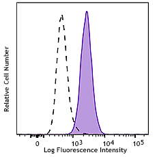
Human peripheral blood granulocytes were stained with CD11b (clone ICRF44) PE (filled histogram) or mouse IgG1, κ PE isotype control (open histogram).
| Cat # | Size | Price | Quantity Check Availability | Save | ||
|---|---|---|---|---|---|---|
| 301305 | 25 tests | $44 | ||||
| 301306 | 100 tests | $110 | ||||
CD11b is a 165-170 kD type I transmembrane glycoprotein also known as αM integrin, Mac-1, CR3, and C3biR. CD11b non-covalently associates with integrin β2 (CD18) and is expressed on granulocytes, monocytes/macrophages, dendritic cells, NK cells, and subsets of T and B cells. CD11b/CD18 is critical for the transendothelial migration of monocytes and neutrophils. It is also involved in granulocyte adhesion, phagocytosis, and neutrophil activation. CD11b/CD18 interacts with ICAM-1 (CD54), ICAM-2 (CD102), ICAM-4, CD14, CD23, heparin, iC3b, fibrinogen, and factor X.
Product DetailsProduct Details
- Verified Reactivity
- Human, Cynomolgus, Rhesus
- Reported Reactivity
- African Green, Baboon, Chimpanzee, Common Marmoset, Pig
- Antibody Type
- Monoclonal
- Host Species
- Mouse
- Formulation
- Phosphate-buffered solution, pH 7.2, containing 0.09% sodium azide and BSA (origin USA)
- Preparation
- The antibody was purified by affinity chromatography, and conjugated with PE under optimal conditions.
- Concentration
- Lot-specific (to obtain lot-specific concentration and expiration, please enter the lot number in our Certificate of Analysis online tool.)
- Storage & Handling
- The antibody solution should be stored undiluted between 2°C and 8°C, and protected from prolonged exposure to light. Do not freeze.
- Application
-
FC - Quality tested
- Recommended Usage
-
Each lot of this antibody is quality control tested by immunofluorescent staining with flow cytometric analysis. For flow cytometric staining, the suggested use of this reagent is 5 µl per million cells in 100 µl staining volume or 5 µl per 100 µl of whole blood.
- Excitation Laser
-
Blue Laser (488 nm)
Green Laser (532 nm)/Yellow-Green Laser (561 nm)
- Application Notes
-
The ICRF44 antibody inhibits heterotypic adhesion of granulocytes in response to fMLP. Additional reported applications (for the relevant formats) include: immunohistochemical staining of acetone-fixed frozen tissue sections, immunofluorescence microscopy5, stimulation of monocytes3, blocking of heterotypic PMN aggregation8, and blocking of granulocyte activation12. This clone was tested in-house and does not work on formalin fixed paraffin-embedded (FFPE) tissue.
The Ultra-LEAF™ purified antibody (Endotoxin < 0.01 EU/µg, Azide-Free, 0.2 µm filtered) is recommended for functional assays (Cat. Nos. 301361 & 301362). -
Application References
(PubMed link indicates BioLegend citation) -
- Knapp W. 1989. Leucocyte Typing IV. Oxford University Press New York.
- Barclay N, et al. 1997. The Leucocyte Antigen Facts Book. Academic Press Inc. San Diego.
- Rezzonico R, et al. 2001. Blood 97:2932. (Stim)
- Marsik C, et al. 2003. Shock 20:493. (FC)
- David A, et al. 2003. J. Leukoc. Biol. 74:551. (IF)
- Charles N, et al. 2010. Nat. Med. 16:701. (FC) PubMed
- Thurlow LR, et al. 2010. Infect. Immun. 128:1128. (FC) PubMed
- Jadhav S, et al. 2001. J. Immunol. 167:5986. (Block)
- Yoshino N, et al. 2000. Exp. Anim. (Tokyo) 49:97. (FC)
- Sestak K, et al. 2007. Vet. Immunol. Immunopathol. 119:21. (FC)
- Wen T, et al. 2014. J Immunol. 192:5481. (FC) PubMed
- Sprong T, et al. 2003. Blood 102:3702. (Block)
- Cash JL, et al. 2013. EMBO Rep. 14:999. (FC) PubMed
- Larsson K, et al. 2015. PNAS. PubMed
- Product Citations
-
- RRID
-
AB_314157 (BioLegend Cat. No. 301305)
AB_314158 (BioLegend Cat. No. 301306)
Antigen Details
- Structure
- Integrin, type I transmembrane glycoprotein, associates with integrin β2 (CD18), 165-170 kD
- Distribution
-
Granulocytes, monocytes/macrophages, dendritic cells, NK cells, subset of T cells, subset of B cells
- Function
- Adhesion, phagocytosis, chemotaxis, neutrophil activation
- Ligand/Receptor
- ICAM-1(CD54), ICAM-2 (CD102), ICAM-4, CD14, CD23, heparin, iC3b, fibrinogen, factor X
- Cell Type
- B cells, Dendritic cells, Granulocytes, Macrophages, Monocytes, Neutrophils, NK cells, T cells, Tregs
- Biology Area
- Cell Adhesion, Cell Biology, Costimulatory Molecules, Immunology, Innate Immunity, Neuroscience, Neuroscience Cell Markers
- Molecular Family
- Adhesion Molecules, CD Molecules
- Antigen References
-
1. Stewart M, et al. 1995. Curr Opin Cell Biol. 7:690.
- Gene ID
- 3684 View all products for this Gene ID
- UniProt
- View information about CD11b on UniProt.org
Related Pages & Pathways
Pathways


Related FAQs
- What type of PE do you use in your conjugates?
- We use R-PE in our conjugates.
Other Formats
View All CD11b Reagents Request Custom ConjugationCustomers Also Purchased
Compare Data Across All Formats
This data display is provided for general comparisons between formats.
Your actual data may vary due to variations in samples, target cells, instruments and their settings, staining conditions, and other factors.
If you need assistance with selecting the best format contact our expert technical support team.
-
APC anti-human CD11b

Human peripheral blood granulocytes stained with ICRF44 APC -
Biotin anti-human CD11b

Human peripheral blood granulocytes stained with biotinylate... -
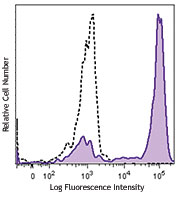
PE anti-human CD11b

Human peripheral blood granulocytes were stained with CD11b ... -
PE/Cyanine5 anti-human CD11b

Human peripheral blood lymphocytes, monocytes, and granulocy... -
Purified anti-human CD11b

Human peripheral blood granulocytes stained with purified IC... -
Pacific Blue™ anti-human CD11b

Human peripheral blood granulocytes stained with ICRF44 Paci... -
Alexa Fluor® 488 anti-human CD11b

Human peripheral blood lymphocytes, monocytes, and granulocy... -
Alexa Fluor® 647 anti-human CD11b

Human peripheral blood granulocytes stained with ICRF44 Alex... -
PE/Cyanine7 anti-human CD11b

Human peripheral blood monocytes stained with ICRF44 PE/Cyan... -
PerCP/Cyanine5.5 anti-human CD11b

Human peripheral blood lymphocytes, monocytes, and granulocy... 
-
Brilliant Violet 421™ anti-human CD11b

Human peripheral blood granulocytes were stained with CD11b ... 
Human neutrophils were fixed with 1% paraformaldehyde (PFA) ... -
Brilliant Violet 570™ anti-human CD11b

Human peripheral blood granulocytes were stained with CD11b ... -
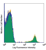
FITC anti-human CD11b

Human peripheral blood granulocytes were stained with CD11b ... -
Brilliant Violet 605™ anti-human CD11b

Human peripheral blood granulocytes were stained with CD11b ... -
Brilliant Violet 510™ anti-human CD11b

Human peripheral blood granulocytes were stained with CD11b ... 
Human neutrophils were fixed with 1% paraformaldehyde (PFA) ... -
Brilliant Violet 650™ anti-human CD11b

Human peripheral blood granulocytes were stained with CD11b ... -
Purified anti-human CD11b (Maxpar® Ready)

Human PBMCs stained with 144Nd-anti-CD11b (ICRF44). T lympho... -
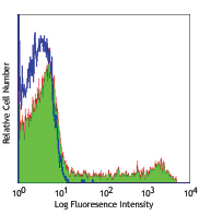
Alexa Fluor® 594 anti-human CD11b

Human peripheral blood mononuclear cells and neutrophil mixe... 
Human peripheral blood granulocytes were stained with CD11b ... -
APC/Cyanine7 anti-human CD11b

Human peripheral blood lymphocytes, monocytes, and granulocy... -
Brilliant Violet 711™ anti-human CD11b

Human peripheral blood granulocytes were stained with CD11b ... -
Brilliant Violet 785™ anti-human CD11b

Human peripheral blood granulocytes were stained with CD11b ... -
PE/Dazzle™ 594 anti-human CD11b

Human peripheral blood granulocytes were stained with CD11b ... -
APC/Fire™ 750 anti-human CD11b

Human peripheral blood granulocytes were stained with CD11b ... -
APC anti-human CD11b

Typical results from human peripheral blood granulocytes sta... -
PE anti-human CD11b

Typical results from human peripheral blood granulocytes sta... -
TotalSeq™-A0161 anti-human CD11b
-
Alexa Fluor® 700 anti-human CD11b

Human peripheral blood lymphocytes, monocytes, and granulocy... -
PE/Cyanine7 anti-human CD11b
Typical results from human peripheral blood monocytes staine... -
TotalSeq™-B0161 anti-human CD11b
-
TotalSeq™-C0161 anti-human CD11b
-
PerCP/Cyanine5.5 anti-human CD11b

Typical results from human peripheral blood granulocytes sta... -
Ultra-LEAF™ Purified anti-human CD11b
Human peripheral blood granulocytes stained with purified IC... -
TotalSeq™-D0161 anti-human CD11b
-
GMP PE/Cyanine7 anti-human CD11b

Typical results from human peripheral blood granulocytes sta... -
Pacific Blue™ anti-human CD11b

Typical results from human peripheral blood granulocytes sta... -
GMP PE anti-human CD11b

Typical results from human peripheral blood granulocytes sta... -
Spark UV™ 387 anti-human CD11b

Human peripheral blood granulocytes were stained with anti-h... -
GMP PerCP/Cyanine5.5 anti-human CD11b

Typical results from human peripheral blood granulocytes sta... -
FITC anti-human CD11b

Typical results from human peripheral blood Granulocytes sta... -
APC/Fire™ 750 anti-human CD11b

Typical results from human peripheral blood granulocytes sta... -
GMP APC anti-human CD11b

Typical results from human peripheral blood granulocytes sta... -
GMP FITC anti-human CD11b

Typical results from human peripheral blood Granulocytes sta... -
GMP APC/Fire™ 750 anti-human CD11b

Typical results from human peripheral blood granulocytes sta... -
Spark Blue™ 515 anti-human CD11b

Human peripheral blood granulocytes were stained with anti-h... 
Human peripheral blood lymphocytes, monocytes, and granulocy... -
Spark Violet™ 500 anti-human CD11b

Human peripheral blood granulocytes were stained with anti-h... -
Spark Red™ 718 anti-human CD11b (Flexi-Fluor™)


Follow Us