 Login/Register


- Clone
- A7R34 (See other available formats)
- Regulatory Status
- RUO
- Other Names
- IL-7 receptor α chain, IL-7Rα
- Isotype
- Rat IgG2a, κ
- Ave. Rating
- Submit a Review
- Product Citations
- publications

-

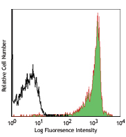
C57BL/6 mouse splenocytes were stained with CD3ε FITC and CD127 (clone A7R34) Brilliant Violet 421™ (top) or rat IgG2a, κ Brilliant Violet 421™ isotype control (bottom). -

CD127 is a 60-90 kD type I transmembrane glycoprotein also known as IL-7 receptor α chain or IL-7Rα. It forms a heterodimer with the common γ chain (γc or CD132) which is shared with the receptors for IL-2, IL-4, IL-9, IL-13, IL-15, and IL-21. CD127 is expressed on immature B cells through early pre-B stage, thymocytes (except CD4/CD8 double positive thymocytes), peripheral T cells, and bone marrow stromal cells. CD127 has been reported to be an useful marker for identifying memory and effector T cells. The ligation of IL-7 with its receptor is important for stimulation of mature and immature T cells as well as immature B cells proliferation and development.
Product DetailsProduct Details
- Verified Reactivity
- Mouse
- Antibody Type
- Monoclonal
- Host Species
- Rat
- Immunogen
- IL-7Ra-IgG1 fusion protein
- Formulation
- Phosphate-buffered solution, pH 7.2, containing 0.09% sodium azide and BSA (origin USA).
- Preparation
- The antibody was purified by affinity chromatography and conjugated with Brilliant Violet 421™ under optimal conditions.
- Concentration
- µg sizes: 0.2 mg/mLµL sizes: lot-specific (to obtain lot-specific concentration and expiration, please enter the lot number in our Certificate of Analysis online tool.)
- Storage & Handling
- The antibody solution should be stored undiluted between 2°C and 8°C, and protected from prolonged exposure to light. Do not freeze.
- Application
-
FC - Quality tested
- Recommended Usage
-
Each lot of this antibody is quality control tested by immunofluorescent staining with flow cytometric analysis. For immunofluorescent staining using the µg size, the suggested use of this reagent is ≤0.5 µg per million cells in 100 µl volume. For immunofluorescent staining using µl sizes, the suggested use of this reagent is 5 µl per million cells in 100 µl staining volume or 5 µl per 100 µl of whole blood. It is recommended that the reagent be titrated for optimal performance for each application.
Brilliant Violet 421™ excites at 405 nm and emits at 421 nm. The standard bandpass filter 450/50 nm is recommended for detection. Brilliant Violet 421™ is a trademark of Sirigen Group Ltd.
Learn more about Brilliant Violet™.
This product is subject to proprietary rights of Sirigen Inc. and is made and sold under license from Sirigen Inc. The purchase of this product conveys to the buyer a non-transferable right to use the purchased product for research purposes only. This product may not be resold or incorporated in any manner into another product for resale. Any use for therapeutics or diagnostics is strictly prohibited. This product is covered by U.S. Patent(s), pending patent applications and foreign equivalents. - Excitation Laser
-
Violet Laser (405 nm)
- Application Notes
-
A7R34 is able to block clone SB/199 binding to IL-7R.
- Additional Product Notes
-
View more applications data for this product in our Scientific Poster Library.
-
Application References
(PubMed link indicates BioLegend citation) - Product Citations
-
- RRID
-
AB_10897948 (BioLegend Cat. No. 135023)
AB_2563103 (BioLegend Cat. No. 135027)
AB_11218800 (BioLegend Cat. No. 135024)
Antigen Details
- Structure
- Type I transmembrane glycoprotein, associate with CD132, 60-90 kD
- Distribution
-
Immature B cells through early pre-B stage, thymocytes (except CD4/CD8 double positive thymocytes), peripheral T cells, bone marrow stromal cells
- Function
- T cell and immature B cell proliferation and development
- Ligand/Receptor
- IL-7
- Cell Type
- B cells, T cells, Thymocytes
- Biology Area
- Immunology
- Molecular Family
- CD Molecules, Cytokine/Chemokine Receptors
- Antigen References
-
1. Sudo T, et al. 1993. P. Natl. Acad. Sci. USA 90:9125.
2. Okuno Y, et al. 2001. P. Natl. Acad. Sci. USA 99:6246.
3. Pillai M, et al. 2004. Leukemia Lymphoma 45:2403. - Gene ID
- 16197 View all products for this Gene ID
- UniProt
- View information about CD127 on UniProt.org
Related Pages & Pathways
Pathways


Related FAQs
- What is the F/P ratio range of our BV421™ format antibody reagents?
-
It is lot-specific. On average it ranges between 2-4.
Other Formats
View All CD127 Reagents Request Custom ConjugationCustomers Also Purchased
Compare Data Across All Formats
This data display is provided for general comparisons between formats.
Your actual data may vary due to variations in samples, target cells, instruments and their settings, staining conditions, and other factors.
If you need assistance with selecting the best format contact our expert technical support team.
-
Purified anti-mouse CD127 (IL-7Rα)

C57BL/6 mouse splenocytes stained with rat IgG2a (RTK2758) P... 
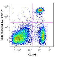
C57BL/6 mouse splenocytes stained with A7R34 PE and CD3 (145... -
FITC anti-mouse CD127 (IL-7Rα)

C57BL/6 splenocytes stained with CD3 (145-2C11) PE and A7R34... 
-
PE anti-mouse CD127 (IL-7Rα)
C57BL/6 mouse splenocytes stained with rat IgG2a (RTK2758) P...
C57BL/6 mouse splenocytes stained with A7R34 PE and CD3 (145... -
APC anti-mouse CD127 (IL-7Rα)

C57BL/6 splenocytes stained with CD3 (145-2C11) PE and A7R34... 
-
PE/Cyanine7 anti-mouse CD127 (IL-7Rα)

C57BL/6 mouse splenocytes stained with CD3ε (clone 1... -
PE/Cyanine5 anti-mouse CD127 (IL-7Rα)

C57BL/6 mouse splenocytes stained with CD3 (clone 145-2C11) ... -
Alexa Fluor® 488 anti-mouse CD127 (IL-7Rα)

C57BL/6 mouse splenocytes stained with CD3 (145-2C11) APC an... 
-
Alexa Fluor® 647 anti-mouse CD127 (IL-7Rα)

Mouse splenocytes stained with CD3 FITC and A7R34 Alexa Fluo... 
-
PerCP/Cyanine5.5 anti-mouse CD127 (IL-7Rα)

C57BL/6 mouse splenocytes stained with CD3 (17A2) PE and A7R... 
-
Biotin anti-mouse CD127 (IL-7Rα)

C57BL/6 mouse splenocytes stained with CD3ε (145-2C11) APC ... 
-
Brilliant Violet 421™ anti-mouse CD127 (IL-7Rα)

C57BL/6 mouse splenocytes were stained with CD3ε FITC and CD... 
-
Brilliant Violet 605™ anti-mouse CD127 (IL-7Rα)

C57BL/6 mouse splenocytes were stained with CD3ε FIT... 
-
Purified anti-mouse CD127 (IL-7Rα) (Maxpar® Ready)

C57BL/6 mouse splenocytes stained with 169Tm-anti-TCRbeta (H... -
PE/Dazzle™ 594 anti-mouse CD127 (IL-7Rα)

C57BL/6 mouse splenocytes were stained with CD3ε FIT... 
-
Brilliant Violet 510™ anti-mouse CD127 (IL-7Rα)

C57BL/6 mouse splenocytes were stained with CD3ε APC... 
-
Brilliant Violet 711™ anti-mouse CD127 (IL-7Rα)

C57BL/6 mouse splenocytes were stained with CD3ε APC... 
-
Brilliant Violet 785™ anti-mouse CD127 (IL-7Rα)

C57BL/6 mouse splenocytes were stained with CD3ε APC... 
-
APC/Cyanine7 anti-mouse CD127 (IL-7Rα)

C57BL/6 mouse splenocytes were stained with CD3 FITC and CD1... -
Brilliant Violet 650™ anti-mouse CD127 (IL-7Rα)

C57BL/6 mouse splenocytes were stained with CD3ε FIT... 
-
TotalSeq™-A0198 anti-mouse CD127 (IL-7Rα)
-
TotalSeq™-C0198 anti-mouse CD127 (IL-7Rα)
-
Ultra-LEAF™ Purified anti-mouse CD127 (IL-7Rα)
-
TotalSeq™-B0198 anti-mouse CD127 (IL-7Rα)


Follow Us