 Login/Register


- Clone
- TER-119 (See other available formats)
- Regulatory Status
- RUO
- Other Names
- Ly-76
- Isotype
- Rat IgG2b, κ
- Ave. Rating
- Submit a Review
- Product Citations
- publications

-

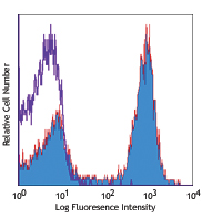
C57BL/6 bone marrow cells were stained with biotinylated TER-119 (filled histogram) or biotinylated rat IgG2b, κ isotype control (open histogram) and then detected with Sav-PE.
| Cat # | Size | Price | Quantity Check Availability | Save | ||
|---|---|---|---|---|---|---|
| 116203 | 50 µg | $47 | ||||
| 116204 | 500 µg | $132 | ||||
The TER-119 antigen is a 52 kD glycophorin A-associated protein, also known as Ly-76. TER-119 is an erythroid-specific antigen expressed on early proerythroblasts to mature erythrocytes, but not on erythroid colony-forming cells (BFU-E, blast-forming unit erythroid, or CFU-E, colony-forming unit erythroid).
Product DetailsProduct Details
- Verified Reactivity
- Mouse
- Antibody Type
- Monoclonal
- Host Species
- Rat
- Immunogen
- Day-14 fetal liver cells from a C57BL/6 mouse
- Formulation
- Phosphate-buffered solution, pH 7.2, containing 0.09% sodium azide.
- Preparation
- The antibody was purified by affinity chromatography, and conjugated with biotin under optimal conditions.
- Concentration
- 0.5 mg/ml
- Storage & Handling
- The antibody solution should be stored undiluted between 2°C and 8°C. Do not freeze.
- Application
-
FC - Quality tested
- Recommended Usage
-
Each lot of this antibody is quality control tested by immunofluorescent staining with flow cytometric analysis. For flow cytometric staining, the suggested use of this reagent is ≤ 0.25 µg per 106 cells in 100 µl volume. It is recommended that the reagent be titrated for optimal performance for each application.
- Application Notes
-
The TER-119 antibody is useful for distinguishing erythrocytes and cells in the erythroid lineage. Additional reported applications (for the relevant formats) include: immunoprecipitation1, Western blotting1, complement-mediated cytotoxicity3, and immunohistochemical staining of acetone-fixed frozen sections and formalin-fixed paraffin-embedded sections. Ultra-LEAF™ purified antibody (Endotoxin < 0.01 EU/µg, Azide-Free, 0.2 µm filtered) is recommended for functional assays (Cat. No. 116253-116258).
-
Application References
(PubMed link indicates BioLegend citation) -
- Kina T, et al. 2000. Br. J. Haematol. 109:280. (IP, WB)
- Vannucchi AM, et al. 2000. Blood 95:2559.
- Maraskovsky E, et al. 1996. J. Exp. Med. 184:1953. (CMCD)
- Grisendi S, et al. 2005. Nature 437:147. (FC)
- Bourdeau A, et al. 2007. Blood 109:4220.
- Chappaz S, et al. 2007. Blood 110:3862. (FC)
- Heuser M, et al. 2007. Blood 110:1639. (FC)
- Gough SM, et al. 2014. Cancer Discov. 4:564. PubMed
- Product Citations
-
- RRID
-
AB_313704 (BioLegend Cat. No. 116203)
AB_313705 (BioLegend Cat. No. 116204)
Antigen Details
- Structure
- Associated with glycophorin A, 52 kD
- Distribution
-
Early proerythroblast to mature erythrocyte, but not BFU-E and CFU-E
- Cell Type
- Erythrocytes
- Biology Area
- Immunology
- Antigen References
-
1. Kina T, et al. 2000. Br. J. Haematol. 109:280.
2. Ikuta K, et al. 1990. Cell 62:863.
3. Osawa M, et al. 1996. Weir's Handbook of Experimental Immunology. Vol. 2 pp. 66.1-66.5. - Gene ID
- 104231 View all products for this Gene ID
- UniProt
- View information about TER-119 on UniProt.org


Related FAQs
- How many biotin molecules are per antibody structure?
- We don't routinely measure the number of biotins with our antibody products but the number of biotin molecules range from 3-6 molecules per antibody.
Other Formats
View All TER-119 Reagents Request Custom ConjugationCustomers Also Purchased
Compare Data Across All Formats
This data display is provided for general comparisons between formats.
Your actual data may vary due to variations in samples, target cells, instruments and their settings, staining conditions, and other factors.
If you need assistance with selecting the best format contact our expert technical support team.
-
APC anti-mouse TER-119/Erythroid Cells

C57BL/6 bone marrow cells stained with TER-119 APC and anti-... -
Biotin anti-mouse TER-119/Erythroid Cells

C57BL/6 bone marrow cells were stained with biotinylated TER... -
FITC anti-mouse TER-119/Erythroid Cells

C57BL/6 bone marrow cells stained with TER-119 FITC and anti... -
PE anti-mouse TER-119/Erythroid Cells

C57BL/6 bone marrow cells stained with TER-119 PE and anti-m... -
PE/Cyanine5 anti-mouse TER-119/Erythroid Cells
-
Purified anti-mouse TER-119/Erythroid Cells
C57BL/6 bone marrow cells stained with TER-119 FITC and CD45... 
Fresh, frozen mouse spleen was stained with purified TER-119... -
Alexa Fluor® 488 anti-mouse TER-119/Erythroid Cells

C57BL/6 mouse bone marrow cells stained with TER119 Alexa Fl... -
Alexa Fluor® 647 anti-mouse TER-119/Erythroid Cells

C57BL/6 bone marrow cells stained with TER-119 Alexa Fluor® ... -
Alexa Fluor® 700 anti-mouse TER-119/Erythroid Cells

C57BL/6 bone marrow stained cell with TER-119 Alexa Fluor&re... -
PE/Cyanine7 anti-mouse TER-119/Erythroid Cells

C57BL/6 mouse bone marrow cells stained with TER119 PE/Cyani... -
APC/Cyanine7 anti-mouse TER-119/Erythroid Cells

C57BL/6 bone marrow cells were stained with TER-119 APC/Cyan... -
PerCP anti-mouse TER-119/Erythroid Cells

C57BL/6 mouse bone marrow cells stained with TER-119 PerCP -
PerCP/Cyanine5.5 anti-mouse TER-119/Erythroid Cells

C57BL/6 mouse bone marrow cells stained with TER-119 PerCP/C... -
Brilliant Violet 421™ anti-mouse TER-119/Erythroid Cells

C57BL/6 splenocytes mixed with red blood cells were stained ... 
-
Pacific Blue™ anti-mouse TER-119/Erythroid Cells

C57BL/6 bone marrow cells stained with Ter-119 Pacific Blue&... -
Brilliant Violet 650™ anti-mouse TER-119/Erythroid Cells

C57BL/6 bone marrow cells were stained with CD45 PE and TER-... -
Brilliant Violet 510™ anti-mouse TER-119/Erythroid Cells

C57BL/6 bone marrow cells were stained with CD45 Pacific Blu... 
-
Brilliant Violet 605™ anti-mouse TER-119/Erythroid Cells

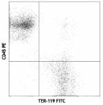
C57BL/6 bone marrow cells were stained with CD45 FITC and TE... 
-
Purified anti-mouse TER-119/Erythroid Cells (Maxpar® Ready)

C57BL/6 mouse splenocytes mixed with red blood cells and sta... -
PE/Dazzle™ 594 anti-mouse TER-119/Erythroid Cells

C57BL/6 bone marrow cells were stained with CD45 APC and TER... 
-
Brilliant Violet 785™ anti-mouse TER-119/Erythroid Cells

C57BL/6 bone marrow cells were stained with CD45 FITC and TE... -
TotalSeq™-A0122 anti-mouse TER-119/Erythroid Cells
-
APC/Fire™ 750 anti-mouse TER-119/Erythroid Cells

C57BL/6 bone marrow cells were stained with CD45 FITC and TE... -
TotalSeq™-B0122 anti-mouse TER-119/Erythroid Cells
-
TotalSeq™-C0122 anti-mouse TER-119/Erythroid Cells
-
Ultra-LEAF™ Purified anti-mouse TER-119/Erythroid Cells
-
Spark Blue™ 550 anti-mouse TER-119/Erythroid Cells

C57BL/6 mouse bone marrow cells stained with CD45 PE and Ter... -
APC/Fire™ 810 anti-mouse TER-119/Erythroid Cells

C57BL/6 bone marrow were stained with CD45 PE and TER-119 (c... -
Spark NIR™ 685 anti-mouse TER-119/Erythroid Cells Antibody

C57BL/6 mouse bone marrow cells were stained with anti-mouse... -
Brilliant Violet 711™ anti-mouse TER-119/Erythroid Cells

C57BL/6 bone marrow cells were stained with anti-mouse CD45 ...


Follow Us