 Login/Register


- Clone
- IT2.2 (See other available formats)
- Regulatory Status
- RUO
- Workshop
- VI CD86.8
- Other Names
- B7-2, B70
- Isotype
- Mouse IgG2b, κ
- Ave. Rating
- Submit a Review
- Product Citations
- publications

-

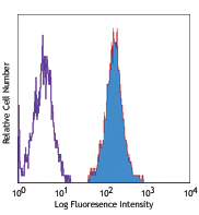
Human peripheral blood monocytes stained with IT2.2 APC
| Cat # | Size | Price | Quantity Check Availability | Save | ||
|---|---|---|---|---|---|---|
| 305411 | 25 tests | $149 | ||||
| 305412 | 100 tests | $314 | ||||
CD86 is an 80 kD immunoglobulin superfamily member also known as B7-2, B70, and Ly-58. CD86 is expressed on activated B and T cells, monocytes/macrophages, dendritic cells, and astrocytes. CD86, along with CD80, is the ligand of CD28 and CD152 (CTLA-4). CD86 is expressed earlier in the immune response than CD80. CD86 has also been shown to be involved in immunoglobulin class-switching and triggering of NK cell-mediated cytotoxicity. CD86 binds to CD28 to transduce costimulatory signals for T cell activation, proliferation, and cytokine production. CD86 can bind to CD152 as well, also known as CTLA-4, to deliver an inhibitory signal to T cells.
Product DetailsProduct Details
- Verified Reactivity
- Human, Cynomolgus, Rhesus
- Reported Reactivity
- African Green, Baboon, Capuchin Monkey, Common Marmoset, Cotton-topped Tamarin, Chimpanzee
- Antibody Type
- Monoclonal
- Host Species
- Mouse
- Formulation
- Phosphate-buffered solution, pH 7.2, containing 0.09% sodium azide and BSA (origin USA)
- Preparation
- The antibody was purified by affinity chromatography, and conjugated with APC under optimal conditions.
- Concentration
- Lot-specific (to obtain lot-specific concentration and expiration, please enter the lot number in our Certificate of Analysis online tool.)
- Storage & Handling
- The antibody solution should be stored undiluted between 2°C and 8°C, and protected from prolonged exposure to light. Do not freeze.
- Application
-
FC - Quality tested
- Recommended Usage
-
Each lot of this antibody is quality control tested by immunofluorescent staining with flow cytometric analysis. For flow cytometric staining, the suggested use of this reagent is 5 µl per million cells in 100 µl staining volume or 5 µl per 100 µl of whole blood.
- Excitation Laser
-
Red Laser (633 nm)
- Application Notes
-
Additional reported applications (for the relevant formats) include: immunohistochemical staining of acetone-fixed frozen tissue sections6, Western blotting3, and blocking of T cell activation2,4,5. The Ultra-LEAF™ purified antibody (Endotoxin < 0.01 EU/µg, Azide-Free, 0.2 µm filtered) is recommended for functional assays (Cat. Nos. 305449 & 305450).
-
Application References
(PubMed link indicates BioLegend citation) -
- Kishimoto T, et al. Eds. 1997. Leucocyte Typing VI. Garland Publishing Inc. London.
- Dieu M. 1998. J. Exp. Med. 188:373. (Block)
- Esser M, et al. 2001. J. Virol. 75:6173. (WB)
- Jeannin P, et al. 1999. J. Immunol. 162:2044. (Block)
- Kapsogeorgou EK, et al. 2001. J. Immunol. 166:3107. (Block)
- Geissmann F, et al. 2001. Blood 97:1241. (IHC)
- Product Citations
-
- RRID
-
AB_493232 (BioLegend Cat. No. 305411)
AB_493231 (BioLegend Cat. No. 305412)
Antigen Details
- Structure
- Ig superfamily, single-chain transmembrane glycoprotein, 80 kD
- Distribution
-
Monocytes/macrophages, activated B cells and T cells, dendritic cells
- Function
- T cell costimulation
- Ligand/Receptor
- CD28, CD152
- Cell Type
- B cells, Dendritic cells, Macrophages, Monocytes, T cells, Tregs
- Biology Area
- Cell Biology, Costimulatory Molecules, Immunology, Neuroscience, Neuroscience Cell Markers
- Molecular Family
- CD Molecules, Immune Checkpoint Receptors
- Antigen References
-
1. Hathcock K, et al. 1996. Adv. Immunol. 62:131.
2. June C, et al. 1994. Immunol. Today 15:321. - Gene ID
- 942 View all products for this Gene ID
- UniProt
- View information about CD86 on UniProt.org
Related Pages & Pathways
Pathways


Other Formats
View All CD86 Reagents Request Custom ConjugationCustomers Also Purchased
Compare Data Across All Formats
This data display is provided for general comparisons between formats.
Your actual data may vary due to variations in samples, target cells, instruments and their settings, staining conditions, and other factors.
If you need assistance with selecting the best format contact our expert technical support team.
-
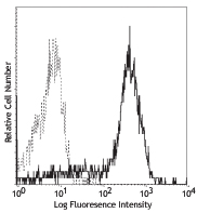
Biotin anti-human CD86

Human peripheral blood monocytes were stained with biotinyla... -
PE anti-human CD86

Human peripheral blood monocytes stained with IT2.2 PE -
PE/Cyanine5 anti-human CD86

Human peripheral blood monocytes were stained with CD86 (clo... -
Purified anti-human CD86

Human peripheral blood monocytes were stained with purified ... -
APC anti-human CD86

Human peripheral blood monocytes stained with IT2.2 APC -
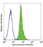
Alexa Fluor® 488 anti-human CD86

Human peripheral blood monocytes with IT2.2 Alexa Fluor®... -
Alexa Fluor® 647 anti-human CD86

Human peripheral blood monocytes stained with IT2.2 Alexa Fl... -
Pacific Blue™ anti-human CD86

Human peripheral blood monocytes stained with IT2.2 Pacific ... -
PE/Cyanine7 anti-human CD86

Human peripheral blood monocytes stained with IT2.2 PE/Cyani... -
PerCP/Cyanine5.5 anti-human CD86

Human peripheral blood monocytes were stained with CD86 (clo... -
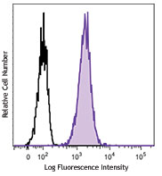
Brilliant Violet 421™ anti-human CD86

Human peripheral blood monocytes were stained with CD86 (clo... -
Brilliant Violet 650™ anti-human CD86

Human peripheral blood monocytes were stained with CD86 (clo... -
Brilliant Violet 605™ anti-human CD86

Human peripheral blood monocytes were stained with CD86 (clo... -
Brilliant Violet 510™ anti-human CD86

Human peripheral blood monocytes were stained with CD86 (clo... -
PE/Dazzle™ 594 anti-human CD86

Human peripheral blood monocytes were stained with CD86 (clo... -
Purified anti-human CD86 (Maxpar® Ready)

Human PBMCs stained with 156Gd-anti-CD86 [B7.2] (IT2.2). Mon... -
Brilliant Violet 711™ anti-human CD86

Human peripheral blood monocytes were stained with CD86 (clo... -
Brilliant Violet 785™ anti-human CD86

Human peripheral blood monocytes were stained with CD86 (clo... -
TotalSeq™-A0006 anti-human CD86
-
TotalSeq™-B0006 anti-human CD86
-
TotalSeq™-C0006 anti-human CD86
-
Ultra-LEAF™ Purified anti-human CD86

Human peripheral blood monocytes were stained with LEAF™ pur... -
KIRAVIA Blue 520™ anti-human CD86

Human peripheral blood monocytes were stained with anti-huma... -
TotalSeq™-D0006 anti-human CD86
-
PE/Fire™ 810 anti-human CD86

Human peripheral monocytes were stained with CD86 (clone IT2... -
PE/Fire™ 640 anti-human CD86

Human peripheral blood cells were blocked with True-Stain Mo... -
PerCP/Fire™ 806 anti-human CD86

Human peripheral blood monocytes were stained with anti-huma... -
PerCP/Fire™ 780 anti-human CD86

Human peripheral blood monocytes were stained with anti-huma... 
Human peripheral blood monocytes were stained with anti-huma...


Follow Us