 Login/Register


- Clone
- W16220A (See other available formats)
- Regulatory Status
- RUO
- Other Names
- Vim, CTRCT30, HEL 113, Epididymis Luminal Protein 113
- Isotype
- Rat IgG2a, κ
- Ave. Rating
- Submit a Review
- Product Citations
- publications

-

NIH/3T3 cells were fixed with 4% paraformaldehyde for 10 minutes, permeabilized with methanol for 6 minutes, and blocked with 5% FBS for 30 minutes. Cells were then intracellularly stained with 2.0 µg/mL (1:250 dilution) Alexa Fluor® 647 rat IgG2a, κ isotype ctrl antibody (Cat. No. 400526) (panel A) or Alexa Fluor® 647 anti-vimentin antibody (clone W16220A) (panel B) overnight at 4°C. Nuclei were counterstained with DAPI, and the image was captured with a 60X objective. -

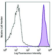
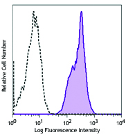
EL-4 cells (positive target) were fixed with fixation buffer (Cat. No. 42080) and permeabilized with True-Phos™ Perm Buffer (Cat. No. 425401), then intracellularly stained with 0.125 µg/test of Alexa Fluor® 647 anti-Vimentin antibody (clone W16220A) (filled histogram) or Alexa Fluor® 647 rat IgG2a, κ isotype ctrl (open histogram). -

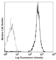
A20 cells (negative target) were fixed with fixation buffer Cat. No. 420801 and permeabilized with True-Phos™ Perm Buffer (Cat. No. 425401), then intracellularly stained with 0.125 µg/test of Alexa Fluor® 647 anti-Vimentin antibody (clone W16220A) (filled histogram) or Alexa Fluor® 647 rat IgG2a, κ isotype ctrl (open histogram).
| Cat # | Size | Price | Quantity Check Availability | Save | ||
|---|---|---|---|---|---|---|
| 699307 | 25 µg | $159 | ||||
| 699308 | 100 µg | $370 | ||||
Vimentin are class-III intermediate filaments found in various non-epithelial cells, especially mesenchymal cells. Vimentin is a widely expressed and highly conserved 54 kD protein that is constitutively expressed in mesenchymal cells, endothelial cells lining blood vessels, renal tubular cells, macrophages, neutrophils, fibroblasts, and leukocytes1,2. Vimentin is used as a marker of mesenchymal cells to distinguish them from epithelial cells3. Increased vimentin expression is frequently used as an EMT marker in cancer4. Autoantibodies to vimentin are commonly found in patients with autoimmune diseases such as Lupus5 and rheumatoid arthritis6, and also found after transplantation7.
Product DetailsProduct Details
- Verified Reactivity
- Mouse
- Antibody Type
- Monoclonal
- Host Species
- Rat
- Immunogen
- Partial mouse Vimentin (224-446 a.a.) recombinant protein expressed in E. coli.
- Formulation
- Phosphate-buffered solution, pH 7.2, containing 0.09% sodium azide
- Preparation
- The antibody was purified by affinity chromatography and conjugated with Alexa Fluor® 647 under optimal conditions.
- Concentration
- 0.5 mg/mL
- Storage & Handling
- The antibody solution should be stored undiluted between 2°C and 8°C, and protected from prolonged exposure to light. Do not freeze.
- Application
-
ICC - Quality tested
ICFC - Verified
SB - Community verified - Recommended Usage
-
Each lot of this antibody is quality control tested by immunocytochemistry. For immunocytochemistry, a concentration range of 1.0 - 5.0 μg/mL is recommended. For intracellular flow cytometric staining, the suggested use of this reagent is ≤ 0.125 µg per million cells in 100 µL volume It is recommended that the reagent be titrated for optimal performance for each application.
* Alexa Fluor® 647 has a maximum emission of 668 nm when it is excited at 633 nm / 635 nm.
Alexa Fluor® and Pacific Blue™ are trademarks of Life Technologies Corporation.
View full statement regarding label licenses - Excitation Laser
-
Red Laser (633 nm)
- Application Notes
-
This antibody does not cross-react with human (in-house tested).
May be weakly reactive with human Vimentin by WB. - Additional Product Notes
-
This product has been verified for IHC-P (Immunohistochemistry - formalin-fixed paraffin-embedded tissues) on the NanoString GeoMx® Digital Spatial Profiler. The GeoMx® enables researchers to perform spatial analysis of protein and RNA targets in FFPE and fresh frozen human and mouse samples. For more information about our spatial biology products and the GeoMx® platform, please visit our spatial biology page.
- RRID
-
AB_2888890 (BioLegend Cat. No. 699307)
AB_2888890 (BioLegend Cat. No. 699308)
Antigen Details
- Structure
- 466 amino acids with a predicted molecular weight of 54 kD.
- Distribution
-
Cytoplasm.
- Function
- Vimentins are class-III intermediate filaments found in various non-epithelial cells, especially mesenchymal cells. Vimentin is attached to the nucleus, endoplasmic reticulum, and mitochondria, either laterally or terminally.
- Interaction
- HCV core protein, LGSN, SYNM, PLEC, SLC6A4, STK33, LARP6, RAB8B, TOR1A, TOR1AIP1, and BCAS3.
- Biology Area
- Cell Biology, Cell Motility/Cytoskeleton/Structure, Neuroscience, Neuroscience Cell Markers
- Molecular Family
- Intermediate Filaments
- Antigen References
-
1. Kidd ME, et al. 2014. Am J Respir Cell Mol Biol. 50:1.
2. Fuchs E, et al. 1994. Annu Rev Biochem. 63:345.
3. Zeisberg M, et al. 2009. J Clin Invest. 119:1429.
4. Scanlon CS, et al. 2013. J Dent Res. 92:114.
5. Thebault S, et al. 2002. J Immunol. 169:4046.
6. Vossenaar ER, et al. 2004. Arthritis Res Ther. 6:R142.
7. Rose ML. 2013. Hum Immunol. 74:1459. - Gene ID
- 22352 View all products for this Gene ID
- UniProt
- View information about Vimentin on UniProt.org
Related Pages & Pathways
Pathways


Related FAQs
- If an antibody clone has been previously successfully used in IBEX in one fluorescent format, will other antibody formats work as well?
-
It’s likely that other fluorophore conjugates to the same antibody clone will also be compatible with IBEX using the same sample fixation procedure. Ultimately a directly conjugated antibody’s utility in fluorescent imaging and IBEX may be specific to the sample and microscope being used in the experiment. Some antibody clone conjugates may perform better than others due to performance differences in non-specific binding, fluorophore brightness, and other biochemical properties unique to that conjugate.
- Will antibodies my lab is already using for fluorescent or chromogenic IHC work in IBEX?
-
Fundamentally, IBEX as a technique that works much in the same way as single antibody panels or single marker IF/IHC. If you’re already successfully using an antibody clone on a sample of interest, it is likely that clone will have utility in IBEX. It is expected some optimization and testing of different antibody fluorophore conjugates will be required to find a suitable format; however, legacy microscopy techniques like chromogenic IHC on fixed or frozen tissue is an excellent place to start looking for useful antibodies.
- Are other fluorophores compatible with IBEX?
-
Over 18 fluorescent formats have been screened for use in IBEX, however, it is likely that other fluorophores are able to be rapidly bleached in IBEX. If a fluorophore format is already suitable for your imaging platform it can be tested for compatibility in IBEX.
- The same antibody works in one tissue type but not another. What is happening?
-
Differences in tissue properties may impact both the ability of an antibody to bind its target specifically and impact the ability of a specific fluorophore conjugate to overcome the background fluorescent signal in a given tissue. Secondary stains, as well as testing multiple fluorescent conjugates of the same clone, may help to troubleshoot challenging targets or tissues. Using a reference control tissue may also give confidence in the specificity of your staining.
- How can I be sure the staining I’m seeing in my tissue is real?
-
In general, best practices for validating an antibody in traditional chromogenic or fluorescent IHC are applicable to IBEX. Please reference the Nature Methods review on antibody based multiplexed imaging for resources on validating antibodies for IBEX.
Other Formats
View All Vimentin Reagents Request Custom Conjugation| Description | Clone | Applications |
|---|---|---|
| Purified anti-Vimentin | W16220A | WB,ICC,ICFC |
| Alexa Fluor® 594 anti-Vimentin | W16220A | ICC |
| Alexa Fluor® 488 anti-Vimentin Antibody | W16220A | ICC,ICFC |
| Alexa Fluor® 647 anti-Vimentin | W16220A | ICC,ICFC,SB |
| PE anti-Vimentin | W16220A | ICFC |
Customers Also Purchased
Compare Data Across All Formats
This data display is provided for general comparisons between formats.
Your actual data may vary due to variations in samples, target cells, instruments and their settings, staining conditions, and other factors.
If you need assistance with selecting the best format contact our expert technical support team.
-
Purified anti-Vimentin

Total cell lysate (15 µg protein) from A20 (negative control... 
NIH3T3 cells were fixed with 4% paraformaldehyde (PFA) for ... 
A20 cells (negative control, open histogram) or EL-4 cells (... -
Alexa Fluor® 594 anti-Vimentin

NIH3T3 cells were fixed with 4% paraformaldehyde (PFA) for 1... -
Alexa Fluor® 488 anti-Vimentin Antibody

NIH/3T3 cells were fixed with 4% paraformaldehyde for 10 min... 
EL4 cells (filled histogram, positive control) and A20 cells... -
Alexa Fluor® 647 anti-Vimentin

NIH/3T3 cells were fixed with 4% paraformaldehyde for 10 min... 
EL-4 cells (positive target) were fixed with fixation buffer... 
A20 cells (negative target) were fixed with fixation buffer ... -
PE anti-Vimentin

EL4 cells (positive control) (filled histogram) and A20 cell...


Follow Us