 Login / Register


- Clone
- 4S.B3 (See other available formats)
- Regulatory Status
- RUO
- Other Names
- Interferon-γ, Immune interferon, Type II interferon, T cell interferon, Macrophage-activating factor (MAF), IFN-g, IFN-gamma
- Isotype
- Mouse IgG1, κ
- Ave. Rating
- Submit a Review
- Product Citations
- publications

-

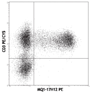
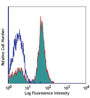
PMA+ionomycin stimulated (6 hours) human peripheral blood lymphocytes (in the presence of monensin) were stained with CD3 PE, then fixed with Fixation Buffer (Cat# 420801), and permeabilized with Permeabilization Wash Buffer (Cat# 421002). Cells were then stained with IFN-γ (clone 4S.B3) APC (top) or mouse IgG1, κ APC isotype control (bottom). -

| Cat # | Size | Price | Quantity Check Availability | Save | ||
|---|---|---|---|---|---|---|
| 502511 | 25 tests | 96€ | ||||
| 502512 | 100 tests | 228€ | ||||
Interferon-γ is a potent multifunctional cytokine which is secreted primarily by activated NK cells and T cells. Originally characterized based on anti-viral activities, IFN-γ also exerts anti-proliferative, immunoregulatory, and proinflammatory activities. IFN-γ can upregulate MHC class I and II antigen expression by antigen-presenting cells.
Product DetailsProduct Details
- Verified Reactivity
- Human
- Reported Reactivity
- Chimpanzee, Baboon, Cynomolgus, Rhesus
- Antibody Type
- Monoclonal
- Host Species
- Mouse
- Immunogen
- Partially purified, native human IFN-γ
- Formulation
- Phosphate-buffered solution, pH 7.2, containing 0.09% sodium azide and BSA (origin USA)
- Preparation
- The antibody was purified by affinity chromatography, and conjugated with APC under optimal conditions.
- Storage & Handling
- The antibody solution should be stored undiluted between 2°C and 8°C, and protected from prolonged exposure to light. Do not freeze.
- Application
-
ICFC - Quality tested
- Recommended Usage
-
Each lot of this antibody is quality control tested by intracellular immunofluorescent staining with flow cytometric analysis. For flow cytometric staining, the suggested use of this reagent is 5 µl per million cells in 100 µl staining volume or 5 µl per 100 µl of whole blood.
- Excitation Laser
-
Red Laser (633 nm)
- Application Notes
-
ELISA or ELISPOT Detection5: The biotinylated 4S.B3 antibody is useful as a detection antibody for a sandwich ELISA or ELISPOT assay, when used in conjunction with purified NIB42 antibody (Cat. No. 502402/502404) or purified MD-1 antibody (Cat. No. 507502/507513) as the capture antibody.
Flow Cytometry3,4,6-8: The fluorochrome-labeled 4S.B3 antibody is useful for intracellular immunofluorescent staining and flow cytometric analysis to identify IFN-γ -producing cells within mixed cell populations.
Additional reported applications (for the relevant formats) include: neutralization1,2, Western blotting, immunohistochemical staining of paraformaldehyde-fixed, saponin-treated tissue sections, and immunocytochemistry. The 4S.B3 antibody can neutralize the bioactivity of natural or recombinant IFN-γ.
Note: For testing human IFN-γ in serum or plasma, BioLegend's ELISA Max™ Sets (Cat. No. 430101 to 430106) are specially developed and recommended. - Application References
-
- Meager A, et al. 1984. J. Interferon Res. 4:619. (Neut)
- Meager A, 1987. Lymphokines and Interferons:A Practical Approach. IRL Press Ltd, Oxford, p. 105. (Neut)
- Sester M, et al. 2002. J. Virol. 76:3748. (ICFC)
- Infante-Duarte C, et al. 2000 J. Immunol. 165:6107. (ICFC)
- Goodier M, et al. 2000. J. Immunol. 165:139. (ELISA)
- Chen H, et al. 2005. J. Immunol. 175:591. (ICFC)
- Smeltz RB, 2007. J. Immunol. 178:4786. (ICFC)
- Iwamoto S, et al. 2007. J. Immunol. 179:1449. (ICFC) PubMed
- Yoshino N, et al. 2000. Exp. Anim. (Tokyo) 49:97. (ICFC)
- Product Citations
-
- RRID
-
AB_315236 (BioLegend Cat. No. 502511)
AB_315237 (BioLegend Cat. No. 502512)
Antigen Details
- Structure
- Cytokine; dimer; 20-25 kD (Mammalian)
- Bioactivity
- Antiviral/antiparasitic activities; inhibits proliferation; enhances MHC class I and II expression on APC
- Cell Sources
- CD8+ and CD4+ T cells, NK cells
- Cell Targets
- T cells, B cells, macrophages, NK cells, endothelial cells, fibroblasts
- Receptors
- IFN-γRα (CDw119) dimerized with IFN-γRβ (AF-1)
- Cell Type
- Tregs
- Biology Area
- Cell Biology, Immunology, Neuroinflammation, Neuroscience
- Molecular Family
- Cytokines/Chemokines
- Antigen References
-
1. Fitzgerald K, et al. Eds. 2001. The Cytokine FactsBook. Academic Press, San Diego.
2. De Maeyer E, et al. 1992. Curr. Opin. Immunol. 4:321.
3. Farrar M, et al. 1993. Annu. Rev. Immunol. 11:571.
4. Gray P, et al. 1987. Lymphokines 13:151. - Regulation
- Upregulated by IL-2, FGF-basic, EGF; downregulated by vitamin D3 or DMN; labile at pH2
- Gene ID
- 3458 View all products for this Gene ID
- UniProt
- View information about IFN-gamma on UniProt.org
Related Pages & Pathways
Pathways


Other Formats
View All IFN-γ Reagents Request Custom ConjugationCustomers Also Purchased
Compare Data Across All Formats
This data display is provided for general comparisons between formats.
Your actual data may vary due to variations in samples, target cells, instruments and their settings, staining conditions, and other factors.
If you need assistance with selecting the best format contact our expert technical support team.
-
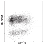
PE anti-human IFN-γ

PMA/Ionomycin-stimulated human PBMCs were stained with CD3 P... -
APC anti-human IFN-γ

PMA+ionomycin stimulated (6 hours) human peripheral blood ly... 
-
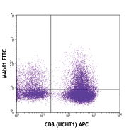
FITC anti-human IFN-γ

PMA/ionomycin-stimulated (6 hours) human peripheral blood ly... 
PMA/ionomycin-stimulated (6 hours) human peripheral blood ly... -
Biotin anti-human IFN-γ

-
Purified anti-human IFN-γ

PMA/Ionomycin-stimulated human PBMCs were stained with CD3 P... 
Western blot analysis of 50 ng recombinant human IFN-γ (lane... -
Alexa Fluor® 488 anti-human IFN-γ

PMA/ionomycin-stimulated (6 hours) human peripheral blood ly... 
PMA/ionomycin-stimulated (6 hours) human peripheral blood ly... -
Alexa Fluor® 647 anti-human IFN-γ

PMA/ionomycin-stimulated (6 hours) human peripheral blood ly... 
PMA/ionomycin-stimulated (6 hours) human peripheral blood ly... -
Alexa Fluor® 700 anti-human IFN-γ

PMA+ionomycin-stimulated (5 hours) human PBMCs surface stain... -
Pacific Blue™ anti-human IFN-γ

PMA+ionomycin-stimulated (6 hours) human peripheral blood ly... -
PerCP/Cyanine5.5 anti-human IFN-γ

PMA+ionomycin-stimulated (6 hours) human peripheral blood ly... -
APC/Cyanine7 anti-human IFN-γ

PMA + Ionomycin (6 hours) stimulated human PBMCs were surfac... -
PE/Cyanine7 anti-human IFN-γ

PMA/ionomycin-stimulated (5 hours) human peripheral blood ly... -
Brilliant Violet 421™ anti-human IFN-γ

Human peripheral blood lymphocytes were stimulated with PMA ... 
-
Brilliant Violet 570™ anti-human IFN-γ

PMA/ionomycin-stimulated (6 hours) human peripheral blood ly... 
-
Brilliant Violet 605™ anti-human IFN-γ

PMA+ionomycin-stimulated (6 hours) human peripheral blood ly... -
Brilliant Violet 650™ anti-human IFN-γ

PMA+ionomycin-stimulated (6 hours) human peripheral blood l... -
Brilliant Violet 711™ anti-human IFN-γ

PMA+ionomycin-stimulated (6 hours) human peripheral blood ly... 
-
Brilliant Violet 785™ anti-human IFN-γ

PMA+ionomycin-stimulated (6 hours) human peripheral blood ly... -
Brilliant Violet 510™ anti-human IFN-γ

PMA/ionomycin-stimulated (6 hours) human peripheral blood ly... 
-
PE/Dazzle™ 594 anti-human IFN-γ

PMA+ Ionomycin-human peripheral blood lymphocytes (in the pr... 
-
APC/Fire™ 750 anti-human IFN-γ

PMA+ionomycin-stimulated (6 hours) human peripheral blood ly... 
-
PerCP anti-human IFN-γ

PMA+ionomycin stimulated (6 hours) human peripheral blood ly... 
-
Brilliant Violet 750™ anti-human IFN-γ

PMA/ionomycin-stimulated (4 hours) human peripheral blood ly... -
KIRAVIA Blue 520™ anti-human IFN-γ Antibody

PMA + Ionomycin (6 hours) stimulated human PBMCs were surfac... -
Spark NIR™ 685 anti-human IFN-γ Antibody

PMA+ ionomycin stimulated (6 hours) human peripheral blood l... -
APC anti-human IFN-γ

Typical result of PMA + Ionomycin with Brefeldin A stimulate... -
Spark Blue™ 515 anti-human IFN-γ

PMA+ Ionomycin (5 hours) stimulated human PBMCs were surface... -
Spark UV™ 387 anti-human IFN-γ

Human peripheral blood lymphocytes were stimulated with Cell... -
Spark PLUS UV™ 395 anti-human IFN-γ

Human peripheral blood lymphocytes were stimulated with PMA/...


Follow Us