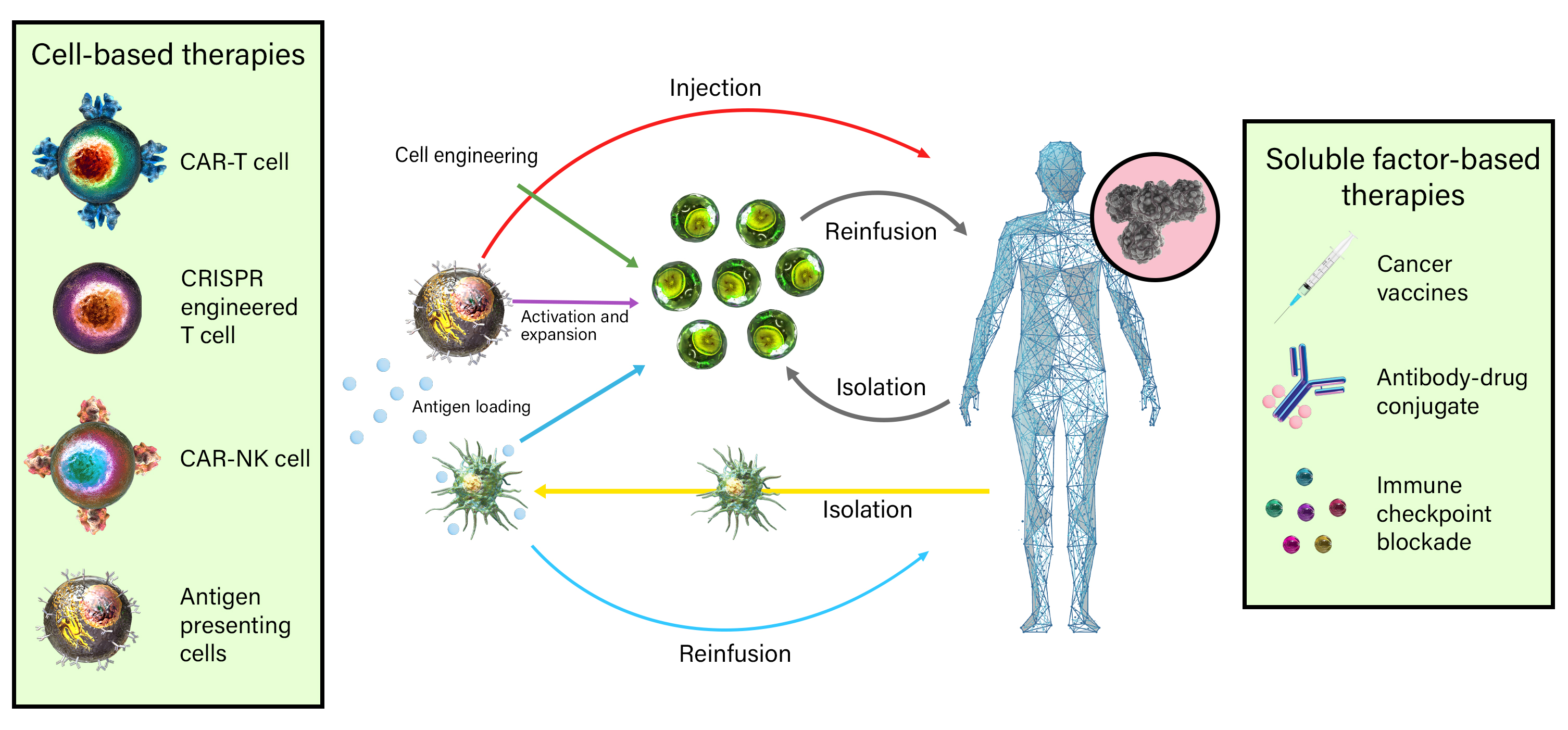
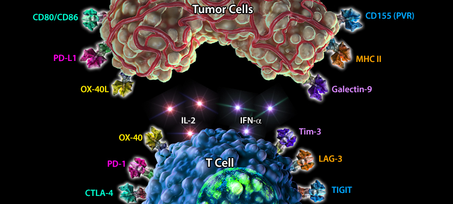
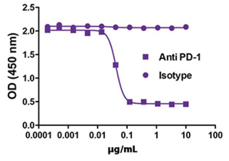
Immuno-oncology is a rapidly moving research area focused on stimulating or helping the immune system fight cancer. Immunotherapies are immune-based treatments for cancer. These include chimeric antigen receptor (CAR) T or Natural Killer (NK) cells engineered to recognize cancer cells, checkpoint inhibitors like anti-PD-1 and anti-CTLA-4 antibodies, cytokines like interleukins and interferons, and therapeutic vaccines that prime the immune system to attack cancer cells. Immunotherapies have shown clinical efficacy in treating cancers, spurring the growth of immunotherapy and immune-oncology research.


Study the roles of cytokines in combating tumor growth with highly stable recombinant proteins, and test strategies for stimulating anti-cancer immune responses with in vivo and ex vivo models with bioactive GoInVivo™ antibodies.

Several forms of immunotherapy-based treatment for cancer are already in clinical use, with many others under development as promising therapeutics. Immunotherapies can be broadly categorized into two strategies: cell-based therapy, where cells are directly injected into patients via stem cells or specific immune cell transplantation, and soluble factor-based therapy, where patients are treated with antibodies and/or other proteins, which boost the patient’s immune system and anti-tumor immunity.

Cell-Based Therapies


Current clinical use and cell-based immunotherapy research is focused around several immune cell types. This includes the transplantation of anti-tumor T cells, such as CAR-T cells, CAR-NK cells, and cancer antigen-primed antigen presenting cells (APC vaccines) 1.

CAR-T Cells


Chimeric antigen receptor T cells (CAR-T cells) are genetically-modified T cells that can be transfused back to patients as a mode of therapy. Unlike traditional T cells carrying T cell receptors (TCRs) that only recognize foreign antigens bound to MHC molecules, CAR-T cells carry artificial antigen-recognizing receptors that do not require an MHC-peptide complex. These receptors are typically introduced into T cells via transfection of the receptor-coding gene2.


CAR-NK Cells


Chimeric antigen receptor NK cells (CAR-NK) are genetically engineered NK cells designed with different chimeric antigen receptors to induce a strong immune response against tumor cells3. NK cells are part of the innate immune system and typically contain granules and cytolytic enzymes that can destroy stressed cells, such as tumor cells. NK cells can be transfected with genes that code for antigen-recognizing receptors and transplanted back to the patient to induce a sustained immune response against cancer cells. These therapies have shown positive results in clinical trials4

KYMRIAH™/Tisagenlecleucel (Novartis) was the first FDA-approved CAR-T cell-based therapy to treat refractory B acute lymphoblastic leukemia (B-ALL). Tisagenlecleucel engineers CAR-T cells to target the B cell marker CD19. Clinical trials have shown statistically significant improvement in survival rates in patients treated with Tisagenlecleucel compared to placebo5. A number of therapies have been approved since for the treatment of a few types of cancers, mainly lymphomas.

A Comparison of CAR-T and CAR-NK Cells


Cancer cells evolve new escape mechanisms. Stay on top of developing technologies including CAR-T and CAR-NK cells with our infographic. Learn about the design, manufacturing, and advantages of these advanced immunotherapies.

Antigen Presenting Cell Vaccines


Antigen Presenting Cell (APC) vaccines rely on the ability of APCs to present tumor-specific antigens as foreign to the host immune system, and mobilize an immune response1. In this cell-based therapy, APCs (primarily monocytes and dendritic cells) are initially harvested from the peripheral blood of patients. The harvested APCs are then cultured with the following:


  • Tumor-specific antigen(s), which is internalized, processed, and displayed by APCs on the cell surface in the form of MHC-tumor peptide complexes.
  • Other soluble factors (such as recombinant proteins) which promote cell maturation and increased immune capabilities.

After maturation and “priming” of the APCs ex vivo, they are transfused back into patients where they will elicit anti-tumor immune responses. After activation, they generate effector and memory T cells to eliminate and surveil tumor recurrence.


One example of an FDA-approved APC vaccine is Sipuleucel-T (Provenge®, Valeant), which is used to treat advanced prostate cancer. In this treatment regimen, monocytes are cultured ex vivo with the tumor antigen prostatic acid phosphatase (PAP) linked to recombinant granulocyte-macrophage colony stimulating factor (GM-CSF) to promote maturation into dendritic cells. The patients then receive multiple injections of the cells. Clinical trials showed statistically significant improvement in survival rates amongst castrate-resistant, metastatic prostate cancer patients treated with Provenge® compared to placebo6.

Soluble Factor-Based Therapies


In order to effectively clear out tumor cells, the immune system needs to be able to generate an appropriate inflammatory response. However, the fact that most of our immune cells contain a fail-safe mechanism to ensure they are not constantly activated is a double-edged sword. There are a number of markers found on both tumor cells and antigen presenting cells that can downregulate or suppress T cells once the corresponding ligand is bound.


Immune Checkpoints


The process of eliciting a T cell-mediated immune response, to eliminate a foreign antigen or an abnormally proliferating cell, is also associated with mechanisms of regulation that ensure a controlled response. These regulatory mechanisms are largely controlled by molecules that researchers have termed ‘immune checkpoint receptors.’ Tumor cells can express high levels of these checkpoint receptors, shutting down immune responses and taking advantage of the lack of inflammation.


Researchers are now looking into how they can prevent T cells from binding these tolerance-inducing ligands. By targeting these molecules with antibodies, we ensure that the T cells stay in an activated state, promoting a sustained response against the cancer cells. For a listing of FDA-approved immune checkpoint inhibitors, visit this webpage by cancer.org.

Although a number of immune checkpoints are being looked at, the most well-studied combinations include PD-1/PD-L1, CTLA-4/CD80 and CD86, LAG-3/MHC II, Tim-3/Galectin 9, and TIGIT/CD155 (PVR) (immune checkpoint combinations have been color-coded based on interaction in the figure above). Additionally, the use of recombinant cytokines, such as IFN-α and IL-2, augment anti-tumor inflammatory responses, and have been used in the clinic as a part of immunotherapeutic regimens to treat various malignancies. The direct injection of molecules involved in inflammation, such as TLR9 and anti-OX40 antibodies, has also shown to be effective in providing long-term anti-tumor protection by promoting and maintaining activation of the immune system.

CTLA-4/CD80 and CD86


CD80 and CD86 (also known as B7-1 and B7-2, respectively) are upregulated on Antigen Presenting Cells (APCs) upon activation of the Toll-Like Receptor (TLR) pathways, and contribute to the activation of T cells by binding to the co-stimulatory molecule CD28. CTLA-4 (or CD152) is upregulated on T cells upon activation and binds to CD80 and CD86 with stronger affinity compared to CD28. This allows preferential complex formation between CD80, CD86 with CTLA-4 instead of CD28. The engagement of the CTLA-4/CD80, CD86 complex results in the attenuation of T cell activation, leading to an immunomodulatory effect. Blockage of CTLA-4 signaling thus allows for prolonged T cell activation, and anti-tumor T cell infiltration6.


CTLA-4 became the first immune checkpoint to be tested for potential cancer treatments. In vivo treatment with anti-CTLA-4 antibodies depleted Tregs, increased CD8+ T cells, and restored T effector function. Treatment with Ipilimumab (anti-CTLA-4 antibody) increased metastatic melanoma patient survival, leading to FDA approval for treatment of melanoma. Pembrolizumab is a possible alternative over Ipilimumab as it has shown improvements in both survival and side effects6.

PD-1/PD-L1


PD-1 is found on activated T cells, Tregs, B cells, NK cells, and myeloid cells. PD-L1 is broadly expressed on hematopoietic and non-hematopoietic cells, including T cells, B cells, NK cells, monocytes, macrophages, granulocytes, and dendritic cells. Activation of PD-1/CD279 signaling via binding to its ligands (PD-L1, PD-L2) leads to downstream signaling that results in cell death. Blockage of PD-1 expressed on activated T cells and its ligands PD-L1 and PD-L2 expressed on tumor cells dampens this cell death process, thus allowing survival of activated T cells that can infiltrate and kill tumor cells7.


Blocking the interaction between PD-1 and PD-L1 showed increased anti-tumor T cell responses in pancreatic carcinomas, B16 melanoma, and CT26 colon carcinoma. Combining CTLA-4 antibody (Ipilimumab) and anti-PD1 antibody (Nivolumab) treatments has resulted in greater anti-tumor responses, with 80% of patients showing tumor regression7,8.

Anti-human PD-1 inhibits the binding of PD-L1. Immobilized PD-1-Fc was pre-incubated with increasing concentrations of anti-human PD-1 (clone EH12.2H7, squares) or isotype control (clone MOPC-21, circles), followed by incubation with a fixed concentration of PD-L1-Fc (1 µg/mL). Clone EH12.2H7 inhibits PD-1/PD-L1 interaction in a dose dependent manner.

OX40 (CD134)/OX40L


OX40 (CD134, TNFRSF4) is a member of the TNF receptor family that is expressed on activated T lymphocytes including Th1, Th2, Th17, and Treg cells. The interaction of OX40 with OX40L results in B cell proliferation and antibody secretion, regulation of primary T cell expansion, and T cell survival by promoting the secretion of IL-29.


Preclinical studies have suggested that OX40, OX40L agonists can boost immune responses that can augment anti-tumor T cell activity, and they are promising targets for immunotherapy. Agonistic anti-OX40 antibodies have been particularly effective when injected, along with a combination of TLR9 and its activating CpG DNA, to act as a “cancer vaccine” that has shown sustained, long-term protection against cancer in preclinical models9.


Recent research highlights the utility of high parameter flow cytometry to address key questions to overcome challenges in T and NK cell therapy for improved treatment10. This includes understanding T and NK cell exhaustion and efforts to improve cytokine release syndrome (CRS) in response to cell therapy.

Flex-T™


Soluble MHC molecules are commonly assembled into tetramers to identify antigen-specific T cells in fields like vaccine development and cancer research. Flex-T™ technology takes MHC monomers and assembles them into tetramers pre-loaded with UV-labile peptides. These peptides in the MHC groove can be swapped out for peptides of interest, allowing antigen-specific T cells to bind through their TCRs.


Learn more about Flex-T™

GMP Solutions


Our GMP recombinant proteins, antibodies, and cell culture reagents are ideal for enrichment, expansion, and quality characterization of cells for bioprocessing needs. The Cell-Vive™ cell culture reagents are advanced formulations: chemically-defined, without animal-derived components, and GMP-grade to yield products that are consistently produced, controlled, and documented.


Our dedicated GMP suite is ISO13485:2016 and MDSAP certified. BioLegend GMP products are manufactured and tested in accordance with USP Chapter 1043, Ancillary Materials for Cell, Gene, and Tissue Engineered Products and Ph. Eur. Chapter 5.2.12.

PBMC-derived T cell culture was activated with anti-human CD3, anti-human CD28, and 200 IU/mL of recombinant human IL-2 using IMDM as basal media plus different additives. On indicated days, cells were quantified.

References

  1. Eggermont, L.J. et al. (2014). Towards efficient cancer immunotherapy: advances in developing artificial antigen-presenting cells. Trends in biotechnology. vol. 32,9: 456-65. doi:10.1016/j.tibtech.2014.06.007. PubMed.
  2. June C.H. (2007). Adoptive T cell therapy for cancer in the clinic. J Clin Invest. 117(6):1466-76. doi: 10.1172/JCI32446. PubMed.
  3. Paul, S. & Girdhari, L. (2017). The Molecular Mechanism of Natural Killer Cells Function and Its Importance in Cancer Immunotherapy. Frontiers in immunology. vol. 8 1124. doi:10.3389/fimmu.2017.01124. PubMed.
  4. Marofi, F. et al. (2021). CAR-NK Cell: A New Paradigm in Tumor Immunotherapy. Frontiers in oncology. vol. 11 673276. doi:10.3389/fonc.2021.673276. PubMed.
  5. Mueller, K.T. et al. (2018). Clinical Pharmacology of Tisagenlecleucel in B-cell Acute Lymphoblastic Leukemia. Clinical cancer research: an official journal of the American Association for Cancer Research vol. 24,24: 6175-6184. doi:10.1158/1078-0432.CCR-18-0758. PubMed.
  6. Kantoff, P. et al. (2010). Sipuleucel-T immunotherapy for castration-resistant prostate cancer. The New England journal of medicine vol. 363,5: 411-22. doi:10.1056/NEJMoa1001294. PubMed.
  7. Hamanishi, J. et al. (2016). PD-1/PD-L1 blockade in cancer treatment: perspectives and issues. International journal of clinical oncology. vol. 21,3: 462-73. doi:10.1007/s10147-016-0959-z. PubMed.
  8. Seidel, J.A. et al. (2018). Anti-PD-1 and Anti-CTLA-4 Therapies in Cancer: Mechanisms of Action, Efficacy, and Limitations. Frontiers in oncology. vol. 8 86. doi:10.3389/fonc.2018.00086. PubMed.
  9. Fu, Y. et al. (2020) Therapeutic strategies for the costimulatory molecule OX40 in T-cell-mediated immunity. Acta pharmaceutica Sinica. B vol. 10,3: 414-433. doi:10.1016/j.apsb.2019.08.010. PubMed.
  10. Nakagawa, R. et al. (2021). High-Dimensional Flow Cytometry Analysis of Regulatory Receptors on Human T Cells, NK Cells, and NKT Cells. Methods in molecular biology. vol. 2194 (2021): 255-290. doi:10.1007/978-1-0716-0849-4_14. PubMed.
ProductsHere

Login / Register
Remember me
Forgot your password? Reset password?
Create an Account