 Login / Register


- Clone
- 10F.9G2 (See other available formats)
- Regulatory Status
- RUO
- Other Names
- B7-H1, PD-L1
- Isotype
- Rat IgG2b, κ
- Ave. Rating
- Submit a Review
- Product Citations
- publications

-

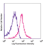
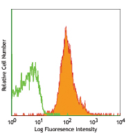
C57/B6 mouse splenocytes were stained with purified anti-CD274 (clone 10F.9G2) (pink line) or purified rat IgG2b, κ isotype control (green line), followed by biotinylated anti-rat IgG and Sav-PE -

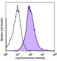
Fresh, frozen mouse spleen was stained with purified CD274 clone 10F.9G2 conjugated and detected with a Cy3 CODEX™ oligonucleotide duplex (red). Samples were counterstained with TCR FITC (greeb). Data generated at Akoya Biosciences, Inc. using the CODEX™ technology.
| Cat # | Size | Price | Quantity Check Availability | Save | ||
|---|---|---|---|---|---|---|
| 124301 | 50 µg | £37 | ||||
| 124302 | 500 µg | £97 | ||||
CD274, also known as B7-H1 or programmed death ligand 1 (PD-L1), is a 40 kD type I transmembrane protein and a member of the B7 family within the immunoglobulin receptor superfamily. It is expressed on T cells, B cells, NK cells, dendritic cells, IFN-γ activated endothelial cells, and monocytes. B7-H1 is one of the ligands of PD-1. The interaction of B7-H1 with PD-1 plays an important role in the inhibition of T cell responses. Other studies have shown that B7-H1 is able to costimulate T cell growth and cytokine production. CD274 is involved in costimulation essential for T cell proliferation and production of IL-10 and IFN-γ, in an IL-2-dependent and a PD-1-independent manner. Its interaction with PD-1 inhibits T cell proliferation and cytokine production.
Product DetailsProduct Details
- Verified Reactivity
- Mouse
- Antibody Type
- Monoclonal
- Host Species
- Rat
- Formulation
- Phosphate-buffered solution, pH 7.2, containing 0.09% sodium azide.
- Preparation
- The antibody was purified by affinity chromatography.
- Concentration
- 0.5 mg/mL
- Storage & Handling
- The antibody solution should be stored undiluted between 2°C and 8°C.
- Application
-
FC - Quality tested
IHC-F - Verified
Block - Reported in the literature, not verified in house - Recommended Usage
-
Each lot of this antibody is quality control tested by immunofluorescent staining with flow cytometric analysis. For flow cytometric staining, the suggested use of this reagent is ≤ 1.0 µg per million cells in 100 µL volume. It is recommended that the reagent be titrated for optimal performance for each application.
- Application Notes
-
Additional reported applications (for the relevant formats) include: immunofluorescence4, blocking6,7,8,9, and immunohistochemistry of acetone-fixed frozen sections4, 11. The LEAF™ purified antibody (Endotoxin <0.1 EU/µg, Azide-Free, 0.2 µm filtered) is recommended for functional assays (Cat. No. 124303). For highly sensitive assays, we recommend Ultra-LEAF™ purified antibody (Cat. No. 124318) with a lower endotoxin limit than standard LEAF™ purified antibodies (Endotoxin <0.01 EU/µg).
-
Application References
(PubMed link indicates BioLegend citation) -
- Maier H, et al. 2007. J. Immunol. 178:2714.
- Meng Q, et al. 2006. Invest. Ophthalmol. Vis. Sci. 47:4444. PubMed
- Scarlett UK, et al. 2012. J Exp Med. 209:495. PubMed
- Grabie N, et al. 2007. Circulation 116:2062. (IF, IHC)
- Paterson AM, et al. 2011. J. Immunol. 187:1097.
- Channappanavar R, et al. 2012. PLoS One 7:e39757. (Block)
- Schreiber HA, et al. 2010. PLoS One 5:e11453. (Block) PubMed
- Muthumani K, et al. 2011. J. Immunol. 187:2932. (Block) PubMed
- Cripps JG, et al. 2010. Hepatology 52:1350. (Block) PubMed
- Murakami R, et al. 2013. PLoS One. 8:73270. PubMed
- Riella LV, et al. 2011. Am. J. Transplant 11:832-40. (IHC)
- Lei GS, et al. 2015. Infect Immun. 83:572. PubMed
- Product Citations
-
- RRID
-
AB_961226 (BioLegend Cat. No. 124301)
AB_961228 (BioLegend Cat. No. 124302)
Antigen Details
- Structure
- 40 kD type I transmembrane protein member of B7 family within the immunoglobulin receptor superfamily
- Distribution
-
T cells, B cells, NK cells, dendritic cells, IFN-γ activated endothelial cells, and monocytes
- Ligand/Receptor
- PD-1 (PDCD1)
- Cell Type
- B cells, Dendritic cells, Endothelial cells, Monocytes, NK cells, T cells
- Biology Area
- Cancer Biomarkers, Costimulatory Molecules, Immunology
- Molecular Family
- Adhesion Molecules, CD Molecules, Immune Checkpoint Receptors
- Antigen References
-
1. Sharpe A, et al. 2007. Nat. Immunol. 8:239.
2. Dong H, et al. 1999. Nat. Med. 5:1365.
3. Freeman G, et al. 2000. J. Exp. Med. 192:1027. - Gene ID
- 60533 View all products for this Gene ID
- UniProt
- View information about CD274 on UniProt.org


Related FAQs
Other Formats
View All CD274 Reagents Request Custom ConjugationCustomers Also Purchased
Compare Data Across All Formats
This data display is provided for general comparisons between formats.
Your actual data may vary due to variations in samples, target cells, instruments and their settings, staining conditions, and other factors.
If you need assistance with selecting the best format contact our expert technical support team.
-
Purified anti-mouse CD274 (B7-H1, PD-L1)

C57/B6 mouse splenocytes were stained with purified anti-CD2... 
Fresh, frozen mouse spleen was stained with purified CD274 c... -
Biotin anti-mouse CD274 (B7-H1, PD-L1)

C57/B6 mouse splenocytes were stained with biotinylated anti... -
PE anti-mouse CD274 (B7-H1, PD-L1)

C57/B6 mouse splenocytes were stained with anti-CD274 (clone... -
Brilliant Violet 421™ anti-mouse CD274 (B7-H1, PD-L1)

C57BL/6 mouse splenocytes were stained with CD274 (clone 10F... -
APC anti-mouse CD274 (B7-H1, PD-L1)

C57/B6 mouse splenocytes were stained with anti-CD274 (clone... -
PE/Cyanine7 anti-mouse CD274 (B7-H1, PD-L1)

C57/B6 mouse splenocytes were stained with anti-CD274 (clone... -
Ultra-LEAF™ Purified anti-mouse CD274 (B7-H1, PD-L1)

C57/B6 mouse splenocytes were stained with Ultra-LEAF™ purif... -
Brilliant Violet 711™ anti-mouse CD274 (B7-H1, PD-L1)

C57BL/6 mouse splenocytes were stained with CD274 (clone 10F... -
Brilliant Violet 605™ anti-mouse CD274 (B7-H1, PD-L1)

C57BL/6 mouse splenocytes were stained with CD274 (clone 10F... -
PE/Dazzle™ 594 anti-mouse CD274 (B7-H1, PD-L1)

C57BL/6 mouse splenocytes were stained with CD274 (clone 10F... -
GoInVivo™ Purified anti-mouse CD274 (B7-H1, PD-L1)

Anti-mouse PD-L1 inhibits the binding of PD-1 to immobilized... -
Brilliant Violet 785™ anti-mouse CD274 (B7-H1, PD-L1)

C57BL/6 mouse splenocytes were stained with CD274 (clone 10F... -
PerCP/Cyanine5.5 anti-mouse CD274 (B7-H1, PD-L1)

C57BL/6 mouse splenocytes were stained with anti-mouse CD274... -
Brilliant Violet 650™ anti-mouse CD274 (B7-H1, PD-L1)

C57BL/6 mouse splenocytes were stained with CD274 (B7-H1, PD... -
PE/Cyanine5 anti-mouse CD274 (B7-H1, PD-L1)

C57BL/6 mouse splenocytes were stained with anti-mouse CD274... -
PE/Fire™ 640 anti-mouse CD274 (B7-H1, PD-L1)

C57BL/6 mouse splenocytes were stained with anti-mouse CD274... -
Spark Red™ 718 anti-mouse CD274 (B7-H1, PD-L1)

C57/B6 mouse splenocytes were stained with anti-mouse CD274 ... -
PE/Fire™ 700 anti-mouse CD274 (B7-H1, PD-L1)

C57BL/6 mouse splenocytes were stained with anti-mouse CD274...


Follow Us