 Login / Register


- Clone
- HA58 (See other available formats)
- Regulatory Status
- RUO
- Workshop
- HCDM listed
- Other Names
- ICAM-1, Ly-47
- Isotype
- Mouse IgG1, κ
- Ave. Rating
- Submit a Review
- Product Citations
- publications

-

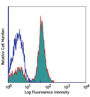
Human peripheral blood lymphocytes were stained with CD54 (clone HA58) PE (filled histogram) or mouse IgG1, κ PE isotype control (open histogram). -

MDA-MB435 breast cancer cell line was stained with anti-human CD54, detected with anti-mouse DyLight™ 649, and nuclear counterstained with DAPI. Images were acquired with a TE300 fluorescence microscope with a 20x objective. Data provided by: Er Liu and John Nolan, La Jolla Bioengineering Institute
| Cat # | Size | Price | Quantity Check Availability | Save | ||
|---|---|---|---|---|---|---|
| 353101 | 25 µg | £37 | ||||
| 353102 | 100 µg | £78 | ||||
CD54 is a 85-110 kD type I transmembrane protein also known as ICAM-1. It is expressed on activated endothelial cells, high endothelial venules, T and B cells, monocytes/macrophages, granulocytes, and dendritic cells. The expression of ICAM-1 can be released from the cell surface. CD54 plays a role in cellular adhesion and is involved in inflammation and leukocyte extravasation. CD54 has also been shown to be the major cellular receptor for rhinovirus. ICAM-1 binds to CD11a/CD18 (LFA-1), CD11b/CD18 (Mac-1), CD11c/CD18 (p150, 95) as well as hyaluronan and fibrinogen.
Product DetailsProduct Details
- Verified Reactivity
- Human
- Antibody Type
- Monoclonal
- Host Species
- Mouse
- Immunogen
- Colonic cancer BM314 cells
- Formulation
- Phosphate-buffered solution, pH 7.2, containing 0.09% sodium azide.
- Preparation
- The antibody was purified by affinity chromatography.
- Concentration
- 0.5 mg/mL
- Storage & Handling
- The antibody solution should be stored undiluted between 2°C and 8°C.
- Application
-
FC - Quality tested
ICC - Verified
IHC - Reported in the literature, not verified in house - Recommended Usage
-
Each lot of this antibody is quality control tested by immunofluorescent staining with flow cytometric analysis. For flow cytometric staining, the suggested use of this reagent is ≤ 0.25 µg per million cells in 100 µL volume. It is recommended that the reagent be titrated for optimal performance for each application.
- Application Notes
-
Clone HA58 recognizes an epitope located in the extracellular D1 domain of CD543. Additional reported applications (for the relevant formats) include: spatial biology (IBEX)4,5.
-
Application References
(PubMed link indicates BioLegend citation) - Product Citations
-
- RRID
-
AB_11204422 (BioLegend Cat. No. 353101)
AB_11204426 (BioLegend Cat. No. 353102)
Antigen Details
- Structure
- Type I membrane protein, Ig superfamily, 85-110 kD
- Distribution
-
Endothelial cells, T cells and B cells, monocytes/macrophages, granulocytes, and dendritic cells
- Cell Type
- B cells, Dendritic cells, Endothelial cells, Granulocytes, Macrophages, Mesenchymal Stem Cells, Monocytes, T cells
- Biology Area
- Cell Adhesion, Cell Biology, Costimulatory Molecules, Immunology, Neuroscience, Neuroscience Cell Markers, Stem Cells
- Molecular Family
- Adhesion Molecules, CD Molecules
- Antigen References
-
1. Voraberger G, et al. 1991. J. Immunol. 147:2777.
2. Staunton DE, et al. 1988. Cell 52:925.
3. Greve JM, et al. 1989. Cell 56:839. - Gene ID
- 3383 View all products for this Gene ID
- UniProt
- View information about CD54 on UniProt.org


Related FAQs
Other Formats
View All CD54 Reagents Request Custom Conjugation| Description | Clone | Applications |
|---|---|---|
| Purified anti-human CD54 | HA58 | FC,ICC,IHC |
| PE anti-human CD54 | HA58 | FC |
| FITC anti-human CD54 | HA58 | FC |
| Pacific Blue™ anti-human CD54 | HA58 | FC |
| APC anti-human CD54 | HA58 | FC |
| Alexa Fluor® 647 anti-human CD54 | HA58 | FC,SB |
| PE/Dazzle™ 594 anti-human CD54 | HA58 | FC |
| APC/Fire™ 750 anti-human CD54 | HA58 | FC |
| PE/Cyanine7 anti-human CD54 | HA58 | FC |
| PerCP/Cyanine5.5 anti-human CD54 | HA58 | FC |
| TotalSeq™-A0217 anti-human CD54 | HA58 | PG |
| Alexa Fluor® 700 anti-human CD54 | HA58 | FC |
| TotalSeq™-C0217 anti-human CD54 | HA58 | PG |
| Alexa Fluor® 488 anti-human CD54 | HA58 | FC |
| Brilliant Violet 421™ anti-human CD54 | HA58 | FC |
| Ultra-LEAF™ Purified anti-human CD54 | HA58 | FC,ICC,IHC-F |
| TotalSeq™-B0217 anti-human CD54 | HA58 | PG |
| TotalSeq™-D0217 anti-human CD54 | HA58 | PG |
| Brilliant Violet 711™ anti-human CD54 | HA58 | FC |
Customers Also Purchased
Compare Data Across All Formats
This data display is provided for general comparisons between formats.
Your actual data may vary due to variations in samples, target cells, instruments and their settings, staining conditions, and other factors.
If you need assistance with selecting the best format contact our expert technical support team.
-
Purified anti-human CD54

Human peripheral blood lymphocytes were stained with CD54 (c... 
MDA-MB435 breast cancer cell line was stained with anti-huma... -
PE anti-human CD54
Human peripheral blood lymphocytes were stained with CD54 (c... -
FITC anti-human CD54

Human peripheral blood lymphocytes were stained with CD54 (c... -
Pacific Blue™ anti-human CD54

Human peripheral blood lymphocytes were stained with CD54 (c... -
APC anti-human CD54

Human peripheral blood lymphocytes were stained with CD54 (c... -
Alexa Fluor® 647 anti-human CD54

Human peripheral blood lymphocytes were stained with CD54 (c... 
Confocal image of human spleen sample acquired using the IBE... 
Confocal image of human liver sample acquired using the IBEX... 
Confocal image of human jejunum sample acquired using the IB... -
PE/Dazzle™ 594 anti-human CD54

Human peripheral blood lymphocytes were stained with CD54 (c... -
APC/Fire™ 750 anti-human CD54

Human peripheral blood lymphocytes were stained with CD54 (c... -
PE/Cyanine7 anti-human CD54

Human peripheral blood lymphocytes were stained with CD54 (c... -
PerCP/Cyanine5.5 anti-human CD54

Human peripheral blood lymphocytes were stained with CD54 (c... -
TotalSeq™-A0217 anti-human CD54
-
Alexa Fluor® 700 anti-human CD54

Human lysed whole blood stained with human CD54 (clone HA58)... -
TotalSeq™-C0217 anti-human CD54
-
Alexa Fluor® 488 anti-human CD54

Human peripheral blood lymphocytes stained with human CD3 Br... -
Brilliant Violet 421™ anti-human CD54

Human peripheral blood lymphocytes were stained with CD54 (c... -
Ultra-LEAF™ Purified anti-human CD54
-
TotalSeq™-B0217 anti-human CD54
-
TotalSeq™-D0217 anti-human CD54
-
Brilliant Violet 711™ anti-human CD54

Human peripheral blood lymphocytes stained with anti-human C...


Follow Us