 Login / Register


- Clone
- W16171A (See other available formats)
- Regulatory Status
- RUO
- Other Names
- H2A.X Variant Histone, H2A Histone Family Member X, Histone H2A.X, Histone H2AX, H2AFX
- Isotype
- Rat IgG2a, κ
- Ave. Rating
- Submit a Review
- Product Citations
- publications

-

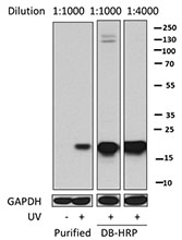
Total cell lysates (15 µg protein) from HeLa (lane 1), HEK293T (lane 2), MCF-7 (lane 3), and NIH/3T3 (lane 4) cells were resolved by 4-20% Tris-Glycine gel electrophoresis, transferred to nitrocellulose, and probed with 1 µg/mL (1:500 dilution) of purified anti-H2A.X antibody (Clone W16171A, left) or a competitor’s clone used at the manufacturer’s recommended dilution. Proteins were visualized by chemiluminescence detection using HRP goat anti-rat IgG (cat #405405) for clone W161171A and HRP donkey anti-rabbit IgG (cat #406401) for the competitor’s clone, each at a 1:3000 dilution. Membranes were then probed with purified anti-GAPDH antibody (lower, cat# 631402) to confirm equal protein loading. Lane M: MW ladder. -

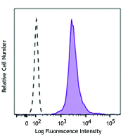
HeLa cells were fixed with 4% paraformaldehyde for 10 minutes, permeabilized with 0.5% Triton X-100 for 10 minutes, and blocked with 5% FBS for 60 minutes. Cells were then intracellularly stained with 1:100 (5 µg/mL), 1:200 (2.0 µg/mL) and 1:500 (1.0 µg/mL) dilutions of either rat IgG or purified anti-H2A.X antibody for two hours at room temperature, followed by incubation with Alexa Fluor® 594 goat anti-rat IgG (cat #405422) at 2.0 µg/mL. Nuclei were counterstained with DAPI, and the image was captured with a 60X objective. -

IHC staining using purified anti-H2A.X antibody (clone W16171A) on formalin-fixed paraffin-embedded human cerebellum tissue. The tissue was incubated with 10 μg/mL of anti-H2A.X antibody overnight at 4°C, followed by incubation with 2.5 μg/mL of Alexa Fluor® 647 goat anti-rat IgG antibody (red) (Cat. No. 405416) for one hour at room temperature. Slide was mounted with ProLong™ Gold Antifade Mountant. Nuclei were counterstained with DAPI (blue, right panel) (Cat. No. 422801). The image was captured with a 40x objective. Scalebar = 50 μM
| Cat # | Size | Price | Quantity Check Availability | Save | ||
|---|---|---|---|---|---|---|
| 600201 | 25 µg | £70 | ||||
| 600202 | 100 µg | £174 | ||||
Histone subunit H2A, along with subunits 2B, 3, and 4, make up the core subunits of the nucleosome octomer. An octomer contains two protomers of each subunit tightly wrapped around a ~147 bp segment of DNA. Histones have integral roles in chromatin integrity, genomic stability, and gene regulation. Post-translational modification of histones in response to certain stimuli results in alterations of nucleosomal positioning relative to DNA. Histone H2A.X is a non-allelic variant of Histone 2A that harbors a C-terminal extension and is essential for checkpoint mediated cell cycle arrest and DNA double-stranded break (DSB) repair in response to both endogenous and exogenous agents, as well as meiotic recombination events and immunoglobulin class switching in lymphocytes. Phosphorylation of C-terminal residue serine 139 by ATM (γ-H2A.X) results in the recruitment of DSB-repair machinery. Phopshorylation of H2A.X is also critical for chromatin fragmentation during apoptosis.
Product DetailsProduct Details
- Verified Reactivity
- Human, Mouse
- Antibody Type
- Monoclonal
- Host Species
- Rat
- Immunogen
- Synthetic human histone H2A.X peptide (127-143) conjugated to KLH.
- Formulation
- Phosphate-buffered solution, pH 7.2, containing 0.09% sodium azide.
- Preparation
- The antibody was purified by affinity chromatography.
- Concentration
- 0.5 mg/ml
- Storage & Handling
- The antibody solution should be stored undiluted between 2°C and 8°C.
- Application
-
WB - Quality tested
ICC, IHC-P - Verified - Recommended Usage
-
Each lot of this antibody is quality control tested by Western blotting. For Western blotting, the suggested use of this reagent is 1.0 - 5.0 µg per ml (1:100-1:500 dilution). For immunocytochemistry, a concentration range of 1.0 - 5.0 μg/ml (1:100-1:500 dilution) is recommended. For immunohistochemistry on formalin-fixed paraffin-embedded tissue sections, a concentration range of 2.0 - 10.0 µg/mL is suggested. It is recommended that the reagent be titrated for optimal performance for each application.
- Application Notes
-
This product is a monoclonal antibody raised against the C-terminus of H2A.X (residues 127-143); BioLegend’s existing antibody against H2A.X (Poly6133, cat# 613302) is a polyclonal antibody which was generated against (partial), N-terminal H2A.X.
This clone is not recommended for ChIP (Chromatin Immunoprecipitation) assays (as determined by in-house testing). - RRID
-
AB_2716198 (BioLegend Cat. No. 600201)
AB_2716199 (BioLegend Cat. No. 600202)
Antigen Details
- Structure
- Histone H2A.X is a 143 amino acid protein with a predicted molecular weight of 15.1 kD.
- Distribution
-
Ubiquitous tissue expression; nuclear localization
- Function
- H2A.X, upon phosphorylation, promotes DNA repair and maintains genomic stability. Important for recombination between immunoglobulin switch regions.
- Interaction
- ATM, MDC1, TP53BP1, BRCA1, MRE11, RAD50, NBN
- Biology Area
- Apoptosis/Tumor Suppressors/Cell Death, Cell Biology, Cell Cycle/DNA Replication, Chromatin Remodeling/Epigenetics, DNA Repair/Replication
- Antigen References
-
- Chen CC, et al. 2017. Proc. Natl. Acad. Sci. 114: 7665.
- Natale F, et al. 2017. Nat. Commun. 8: 15760.
- Bhargava R, et al. 2017. Proc. Natl. Acad. Sci. 114: 728.
- Weyemi U, et al. 2016. Nat. Commun. 7: 10711.
- Rezaeian AH, et al. 2017. Nat. Cell. Biol. 19: 38.
- Horn S, et al. 2015. Biochim. Biophys. Acta. 1853: 2199.
- Reina-San-Martin B, et al. 2003. J. Exp. Med. 197:1767
- Celeste A, et al. 2002. Science 296:922.
- Mannironi C, et al.1989. Nucleic Acids Res. 17:9113.
- Gene ID
- 3014 View all products for this Gene ID
- UniProt
- View information about H2A.X on UniProt.org

Related FAQs
Other Formats
View All H2A.X Reagents Request Custom Conjugation| Description | Clone | Applications |
|---|---|---|
| Purified anti-H2A.X | W16171A | WB,ICC,IHC-P |
| Alexa Fluor® 594 anti-H2A.X | W16171A | ICC |
| Alexa Fluor® 647 anti-H2A.X | W16171A | ICC |
| Alexa Fluor® 488 anti-H2A.X | W16171A | ICC |
Customers Also Purchased
Compare Data Across All Formats
This data display is provided for general comparisons between formats.
Your actual data may vary due to variations in samples, target cells, instruments and their settings, staining conditions, and other factors.
If you need assistance with selecting the best format contact our expert technical support team.
-
Purified anti-H2A.X

Total cell lysates (15 µg protein) from HeLa (lane 1), HEK29... 
HeLa cells were fixed with 4% paraformaldehyde for 10 minute... 
IHC staining using purified anti-H2A.X antibody (clone W1617... -
Alexa Fluor® 594 anti-H2A.X

HeLa cells were fixed with 4% paraformaldehyde for 10 minute... -
Alexa Fluor® 647 anti-H2A.X

HeLa cells were fixed with 4% paraformaldehyde for 10 minute... -
Alexa Fluor® 488 anti-H2A.X

HeLa cells were fixed with 4% paraformaldehyde for 10 minute...


Follow Us