 Login / Register


- Clone
- GL3 (See other available formats)
- Regulatory Status
- RUO
- Other Names
- T cell receptor γ/δ
- Isotype
- Armenian Hamster IgG
- Ave. Rating
- Submit a Review
- Product Citations
- publications

-

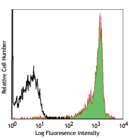
C57BL/6 splenocytes stained with CD3e (145-2C11) FITC and GL3 PerCP/Cyanine5.5 -

C57BL/6 splenocytes stained with CD3e (145-2C11) FITC and Armenian Hamster IgG PerCP/Cyanine5.5 isotype control
| Cat # | Size | Price | Quantity Check Availability | Save | ||
|---|---|---|---|---|---|---|
| 118117 | 25 µg | £81 | ||||
| 118118 | 100 µg | £221 | ||||
T cell receptor (TCR) is a heterodimer consisting of an α and a β chain (TCR α/β) or a γ and a δ chain (TCR γ/δ). TCR γ/δ belongs to the immunoglobulin superfamily, which is involved in the recognition of certain bacterial and tumor antigens bound to MHC class I. γ/δ TCR associates with CD3 and is expressed on a T cell subset found in the thymus, the intestinal epithelium, and the peripheral lymphoid tissues and peritoneum. Most γ/δ T cells are CD4-/CD8- although some are CD8+. T cells expressing the γ/δ TCR have been shown to play a role in oral tolerance, tumor-associated tolerance, and autoimmune disease. It has been reported that γ/δ T cells also play a principal role in antigen presentation.
Product DetailsProduct Details
- Verified Reactivity
- Mouse
- Antibody Type
- Monoclonal
- Host Species
- Armenian Hamster
- Immunogen
- C57BL/6J intraepithelial lymphocytes
- Formulation
- Phosphate-buffered solution, pH 7.2, containing 0.09% sodium azide.
- Preparation
- The antibody was purified by affinity chromatography and conjugated with PerCP/Cyanine5.5 under optimal conditions.
- Concentration
- 0.2 mg/ml
- Storage & Handling
- The antibody solution should be stored undiluted between 2°C and 8°C, and protected from prolonged exposure to light. Do not freeze.
- Application
-
FC - Quality tested
- Recommended Usage
-
Each lot of this antibody is quality control tested by immunofluorescent staining with flow cytometric analysis. For flow cytometric staining, the suggested use of this reagent is ≤1.0 µg per million cells in 100 µl volume. It is recommended that the reagent be titrated for optimal performance for each application.
* PerCP/Cyanine5.5 has a maximum absorption of 482 nm and a maximum emission of 690 nm. - Application Notes
-
The GL3 antibody has been shown to be useful in identifying γ/δ T cells by flow cytometry and immunohistochemistry and depleting γ/δ T cells in vivo. Additional reported applications (for the relevant formats) include: immunoprecipitation1, immunohistochemistry of acetone-fixed frozen sections2,6, and in vivo depletion of γ/δ T cells3-5.
- Additional Product Notes
- BioLegend is in the process of converting the name PerCP/Cy5.5 to PerCP/Cyanine5.5. The dye molecule remains the same, so you should expect the same quality and performance from our PerCP/Cyanine5.5 products. Contact Technical Service if you have any questions.
-
Application References
(PubMed link indicates BioLegend citation) -
- Goodman T, et al. 1989. J. Exp. Med. 170:1569. (FC, IP)
- Cardona AE, et al. 2003. Infect. Immun. 71:2634. (IHC)
- Kapp JA, et al. 2004. Immunology 111:155. (Deplete)
- Skelsey ME, et al. 2001. J. Immunol. 166:4327. (Deplete)
- Ke Y, et al. 1997. J. Immunol. 158:3610. (Deplete)
- Podd BS, et al. 2006. J. Immunol. 176:6532. (IHC)
- Kasten KR, et al. 2010. Infect. Immun. 78:4714. (FC) PubMed
- Stadanlick JE, et al. 2011. J. Immunol. 187:664. PubMed
- Van Belle AB, et al. 2012. J. Immunol. 188:462. PubMed
- Product Citations
-
- RRID
-
AB_10612572 (BioLegend Cat. No. 118117)
AB_10612756 (BioLegend Cat. No. 118118)
Antigen Details
- Structure
- Ig superfamily, associates with CD3 complex.
- Distribution
-
T cell subset in thymus, intestinal epithelium, peripheral lymphoid tissues and peritoneum, most γ/δ T cells are CD4-/CD8-, some are CD8+.
- Function
- Antigen recognition; γ/δ T cells are thought to play a role in tolerance.
- Ligand/Receptor
- Some bacterial or tumor antigens bound to MHC class I.
- Cell Type
- Epithelial cells, T cells, Tregs
- Biology Area
- Adaptive Immunity, Immunology
- Molecular Family
- TCRs
- Antigen References
-
- Skarstein K, et al. 1995. Immunology. 81:497.
- Harrison LC, et al. 1996. J Exp Med. 184:2167.
- Wildner G, et al. 1996. Eur J Immunol. 26:2140.
- Brandes M, et al. 2005. Science. 309:264.
- Gene ID
- 110066 View all products for this Gene ID 110067 View all products for this Gene ID
- UniProt
- View information about TCR gamma/delta on UniProt.org
Related Pages & Pathways
Pathways


Related FAQs
- How stable is PerCP/Cyanine5.5 tandem as compared to PerCP alone?
-
PerCP/Cyanine5.5 is quite photostable and also better than PerCP alone in withstanding fixation.
Other Formats
View All TCR γ/δ Reagents Request Custom Conjugation| Description | Clone | Applications |
|---|---|---|
| Brilliant Violet 421™ anti-mouse TCR γ/δ | GL3 | FC |
| Purified anti-mouse TCR γ/δ | GL3 | FC,IHC-F,IP |
| Biotin anti-mouse TCR γ/δ | GL3 | FC |
| FITC anti-mouse TCR γ/δ | GL3 | FC |
| PE anti-mouse TCR γ/δ | GL3 | FC,SB |
| APC Anti-mouse TCR γ/δ | GL3 | FC |
| PerCP/Cyanine5.5 anti-mouse TCR γ/δ | GL3 | FC |
| PE/Cyanine7 anti-mouse TCR γ/δ | GL3 | FC |
| Alexa Fluor® 488 anti-mouse TCR γ/δ | GL3 | FC |
| Brilliant Violet 605™ anti-mouse TCR γ/δ | GL3 | FC |
| Brilliant Violet 510™ anti-mouse TCR γ/δ | GL3 | FC |
| Alexa Fluor® 647 anti-mouse TCR γ/δ | GL3 | FC |
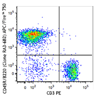
| APC/Fire™ 750 anti-mouse TCR γ/δ | GL3 | FC |
| TotalSeq™-A0121 anti-mouse TCR γ/δ | GL3 | PG |
| Ultra-LEAF™ Purified anti-mouse TCR γ/δ | GL3 | FC,IHC-F,IP |
| TotalSeq™-C0121 anti-mouse TCR γ/δ | GL3 | PG |
| APC/Cyanine7 anti-mouse TCR γ/δ | GL3 | FC |
| TotalSeq™-B0121 anti-mouse TCR γ/δ | GL3 | PG |
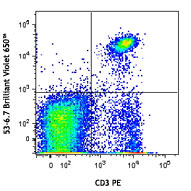
| Brilliant Violet 650™ anti-mouse TCR γ/δ | GL3 | FC |
| Brilliant Violet 711™ anti-mouse TCR γ/δ | GL3 | FC |
| Spark Red™ 718 anti-mouse TCR γ/δ (Flexi-Fluor™) | GL3 | FC |
Customers Also Purchased
Compare Data Across All Formats
This data display is provided for general comparisons between formats.
Your actual data may vary due to variations in samples, target cells, instruments and their settings, staining conditions, and other factors.
If you need assistance with selecting the best format contact our expert technical support team.
-
Brilliant Violet 421™ anti-mouse TCR γ/δ


-
Purified anti-mouse TCR γ/δ
-
Biotin anti-mouse TCR γ/δ

C57BL/6 lymph node cells stained with biotinylated GL3 and C... -
FITC anti-mouse TCR γ/δ

C57BL/6 lymph node cells stained with GL3 FITC and CD3 (145-... -
PE anti-mouse TCR γ/δ

C57BL/6 splenocytes stained with CD3 (145-2C11) APC and GL3 ... 
Confocal image of C57BL/6 mouse thymus sample acquired using... -
APC Anti-mouse TCR γ/δ

C57BL/6 mouse lymph node cells stained with CD3 (145-2C11) P... 
C57BL/6 mouse lymph node cells stained with CD3 (145-2C11) P... -
PerCP/Cyanine5.5 anti-mouse TCR γ/δ

C57BL/6 splenocytes stained with CD3e (145-2C11) FITC and GL... 
C57BL/6 splenocytes stained with CD3e (145-2C11) FITC and Ar... -
PE/Cyanine7 anti-mouse TCR γ/δ

C57BL/6 mouse splenocytes were stained with CD3 APC and TCR ... -
Alexa Fluor® 488 anti-mouse TCR γ/δ

C57BL/6 mouse splenocytes were stained with CD3 Alexa Fluor®... 
-
Brilliant Violet 605™ anti-mouse TCR γ/δ

C57BL/6 mouse splenocytes were stained with CD3ε APC and TC... -
Brilliant Violet 510™ anti-mouse TCR γ/δ

C57BL/6 mouse splenocytes were stained with CD3 APC and TCR ... 
-
Alexa Fluor® 647 anti-mouse TCR γ/δ

C57BL/6 mouse splenocytes were stained with CD3 FITC and TCR... 
-
APC/Fire™ 750 anti-mouse TCR γ/δ

C57BL/6 mouse splenocytes were stained with CD3 PE and and T... 
-
TotalSeq™-A0121 anti-mouse TCR γ/δ
-
Ultra-LEAF™ Purified anti-mouse TCR γ/δ
-
TotalSeq™-C0121 anti-mouse TCR γ/δ
-
APC/Cyanine7 anti-mouse TCR γ/δ

C57BL/6 mouse splenocytes were stained with anti-mouse CD3 F... -
TotalSeq™-B0121 anti-mouse TCR γ/δ
-
Brilliant Violet 650™ anti-mouse TCR γ/δ

C57BL/6 mouse splenocytes were surface stained with anti-mou... -
Brilliant Violet 711™ anti-mouse TCR γ/δ

C57BL/6 splenocytes were stained with anti-mouse CD3 FITC an... -
Spark Red™ 718 anti-mouse TCR γ/δ (Flexi-Fluor™)


Follow Us