 Login / Register


- Clone
- H57-597 (See other available formats)
- Regulatory Status
- RUO
- Other Names
- TCR-β chain, TCR-β, β-TCR
- Isotype
- Armenian Hamster IgG
- Ave. Rating
- Submit a Review
- Product Citations
- publications

-

C57BL/6 mouse splenocytes stained with H57-597 PE/Cyanine7
| Cat # | Size | Price | Quantity Check Availability | Save | ||
|---|---|---|---|---|---|---|
| 109221 | 25 µg | £65 | ||||
| 109222 | 100 µg | £151 | ||||
T cell receptor (TCR) is a heterodimer consisting of an α and a β chain (TCR α/β) or a γ and a δ chain (TCR γ/δ). TCR-β is a member of the immunoglobulin superfamily and a component of the CD3/TCR complex (along with TCR-α). It is expressed on α/β TCR-bearing T cells and thymocytes. The CD3/TCR complex plays a key role in antigen recognition, signal transduction, and T cell activation.
Product DetailsProduct Details
- Verified Reactivity
- Mouse
- Antibody Type
- Monoclonal
- Host Species
- Armenian Hamster
- Immunogen
- Affinity purified TCR from mouse DO-11.10 cells
- Formulation
- Phosphate-buffered solution, pH 7.2, containing 0.09% sodium azide.
- Preparation
- The antibody was purified by affinity chromatography, and conjugated with PE/Cyanine7 under optimal conditions.
- Concentration
- 0.2 mg/ml
- Storage & Handling
- The antibody solution should be stored undiluted between 2°C and 8°C, and protected from prolonged exposure to light. Do not freeze.
- Application
-
FC - Quality tested
- Recommended Usage
-
Each lot of this antibody is quality control tested by immunofluorescent staining with flow cytometric analysis. For flow cytometric staining, the suggested use of this reagent is ≤ 0.25 µg per 106 cells in 100 µl volume. It is recommended that the reagent be titrated for optimal performance for each application.
- Excitation Laser
-
Blue Laser (488 nm)
Green Laser (532 nm)/Yellow-Green Laser (561 nm)
- Application Notes
-
H57-597 is a hamster mAb directed to an epitope of the C region of TCR ß chain12. The H57-597 antibody does not cross-react with ?/d TCR-bearing T cells. Immobilized or soluble H57-597 antibody can activate a/ß TCR-bearing T cells. Additional reported applications (for the relevant formats) for this antibody include: immunoprecipitation2, in vitro stimulation2,3, in vivo depletion4-6, and immunohistochemical staining of acetone-fixed frozen sections7,8,9. The Ultra-LEAF™ purified antibody (Endotoxin <0.01 EU/µg, Azide-Free, 0.2 µm filtered) is recommended for functional assays (Cat. No. 109253-109258).
- Additional Product Notes
- BioLegend is in the process of converting the name PE/Cy7 to PE/Cyanine7. The dye molecule remains the same, so you should expect the same quality and performance from our PE/Cyanine7 products. Please contact Technical Service if you have any questions.
-
Application References
(PubMed link indicates BioLegend citation) -
- Gascoigne NJ. 1990. J. Biol. Chem. 265:9296.
- Kruisbeek A, et al. 1991. In Current Protocols in Immunology. pp. 3.12.1. (Costim IP)
- Davenport C, et al. 1995. J. Immunol. 155:3742. (Costim)
- Drobyski W, et al. 1996. Blood 87:5355. (Deplete)
- Kummer U, et al. 2001. Immunol. Lett. 75:153. (Deplete)
- van der Heyde HC, et al. 1995. J. Immunol. 154:3985. (Deplete)
- Tomita K, et al. 1999. Genes Dev. 13:1203. (IHC)
- Podd BS, et al. 2006. J. Immunol. 176:6532. (IHC)
- Ponomarev ED, et al. 2007. J. Immunol. 178:39. (IHC)
- Chappaz S, et al. 2007. Blood doi:10.1182/blood-2007-02-074245. (FC) PubMed
- Tsukumo S, et al. 2006. J.Immunol. 177:8365. (FC) PubMed
- Grégoire C, et al. 1991. Proc. Natl. Acad. Sci USA 88:8077.
- Product Citations
-
- RRID
-
AB_893627 (BioLegend Cat. No. 109221)
AB_893625 (BioLegend Cat. No. 109222)
Antigen Details
- Structure
- Ig superfamily, CD3/TCR complex with CD3 and TCR α subunit
- Distribution
-
Majority of T cells and thymocytes (correlated to differentiation)
- Function
- Antigen recognition, T cell activation
- Ligand/Receptor
- Peptide bound-MHC class I and II
- Antigen References
-
1. Davis MM, et al. 1998. Ann. Rev. Immunol. 16:523.
2. Huppa JB, et al. 2003. Nat. Immunol. 4:749.
3. Kubo R, et al. 1989. J. Immunol. 142:2736. - Gene ID
- 21577 View all products for this Gene ID
- UniProt
- View information about TCR beta chain on UniProt.org


Related FAQs
Other Formats
View All TCR β chain Reagents Request Custom ConjugationCustomers Also Purchased
Compare Data Across All Formats
This data display is provided for general comparisons between formats.
Your actual data may vary due to variations in samples, target cells, instruments and their settings, staining conditions, and other factors.
If you need assistance with selecting the best format contact our expert technical support team.
-
APC anti-mouse TCR β chain

C57BL/6 mouse splenocytes stained with H57-597 APC -
Biotin anti-mouse TCR β chain

C57BL/6 mouse splenocytes stained with biotinylated H57-597,... 
C57BL/6 mouse frozen spleen section was fixed with 4% parafo... -
FITC anti-mouse TCR β chain

C57BL/6 mouse splenocytes stained with H57-597 FITC -
PE anti-mouse TCR β chain

C57BL/6 CD3+ splenocytes stained with H57-597 PE and TCRγ/δ ... -
PE/Cyanine5 anti-mouse TCR β chain

C57BL/6 mouse splenocytes stained with H57-597 PE/Cyanine5. -
Purified anti-mouse TCR β chain

C57L/6 splenocytes stained with H57-597 purified, followed b... 
Fresh, frozen mouse spleen was stained with purified TCR β c... -
Alexa Fluor® 488 anti-mouse TCR β chain

C57BL/6 mouse splenocytes stained with H57-597 Alexa Fluor® ... -
Alexa Fluor® 647 anti-mouse TCR β chain

C57BL/6 splenocytes stained with H57-597 Alexa Fluor® 647 
C57BL/6 mouse frozen spleen section was fixed with 4% parafo... -
APC/Cyanine7 anti-mouse TCR β chain

C57BL/6 mouse splenocytes were stained with TCR β (clon... -
PE/Cyanine7 anti-mouse TCR β chain

C57BL/6 mouse splenocytes stained with H57-597 PE/Cyanine7 -
Alexa Fluor® 700 anti-mouse TCR β chain

C57BL/6 mouse splenocytes stained with H57-597 Alexa Fluor&r... -
Pacific Blue™ anti-mouse TCR β chain

C57BL/6 splenocytes stained with H57-597 Pacific Blue™ -
Brilliant Violet 421™ anti-mouse TCR β chain

C57BL/6 mouse splenocytes were stained with TCR β (clone H57... -
PerCP/Cyanine5.5 anti-mouse TCR β chain

C57BL/6 splenocytes were stained with CD3e APC and TCR ß cha... -
Brilliant Violet 570™ anti-mouse TCR β chain

C57BL/6 mouse splenocytes were stained with TCR β (clon... -
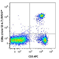
Brilliant Violet 510™ anti-mouse TCR β chain

C57BL/6 mouse splenocytes were stained with TCR β (clone H57... -
Purified anti-mouse TCR β chain (Maxpar® Ready)

Mouse splenocytes stained with 143Nd-anti-TCRβ (H57-597) and... -
Alexa Fluor® 594 anti-mouse TCR β chain

C57BL/6 mouse frozen lymph node sections were fixed with 4% ... 
C57BL/6 mouse splenocytes were stained with TCR β (clone H57... -
PE/Dazzle™ 594 anti-mouse TCR β chain

C57BL/6 mouse splenocytes were stained with TCR-β (clon... -
Brilliant Violet 605™ anti-mouse TCR β chain

C57BL/6 mouse splenocytes were stained with TCR β (clon... -
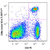
Brilliant Violet 711™ anti-mouse TCR β chain

C57BL/6 mouse splenocytes were stained with TCR β (clon... -
APC/Fire™ 750 anti-mouse TCR β chain

C57BL/6 splenocytes were stained with TCR γ/δ PE... 
-
TotalSeq™-A0120 anti-mouse TCR β chain
-
Brilliant Violet 785™ anti-mouse TCR β chain

C57BL/6 splenocytes were stained with CD3 APC and anti-mouse... -
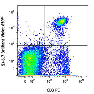
Brilliant Violet 650™ anti-mouse TCR β chain

C57BL/6 Splenocytes were stained with CD3 APC and anti-mouse... -
Ultra-LEAF™ Purified anti-mouse TCR β chain
C57BL/6 CD3+ splenocytes stained with H57-597 PE ... -
TotalSeq™-C0120 anti-mouse TCR β chain
-
TotalSeq™-B0120 anti-mouse TCR β chain
-
Spark Red™ 718 anti-mouse TCR β chain (Flexi-Fluor™)


Follow Us