 Login / Register


- Clone
- 9C4 (See other available formats)
- Regulatory Status
- RUO
- Other Names
- Ep-CAM, tumor associated calcium signal transducer 1, epithelial cell surface antigen, epithelial glycoprotein 2, EGP2, adenocarcinoma associated antigen, TROP1
- Isotype
- Mouse IgG2b, κ
- Ave. Rating
- Submit a Review
- Product Citations
- publications

-

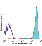
HT-29 cells were stained with CD326 (EpCAM) PE/Cyanine7 (filled histogram) or mouse IgG2b, κ PE/Cyanine7 isotype control (open histogram).
| Cat # | Size | Price | Quantity Check Availability | Save | ||
|---|---|---|---|---|---|---|
| 324221 | 25 tests | £121 | ||||
| 324222 | 100 tests | £262 | ||||
CD326 is also known as Ep-CAM, tumor associated calcium signal transducer 1, epithelial cell surface antigen, epithelial glycoprotein 2, EGP2, adenocarcinoma associated antigen, and TROP1. CD326 is a type I transmembrane protein containing six disulfide bridges and one THYRO domain. This cell surface glycosylated 40 kD protein is highly expressed in bone marrow, colon, lung, and most normal epithelial cells and is expressed on carcinomas of gastrointestinal origin. Recently, it has been reported that CD326 expression occurs during the early steps of erythrogenesis. CD326 functions as a homotypic calcium-independent cell adhesion molecule and is believed to be involved in carcinogenesis by its ability to induce genes involved in cellular metabolism and proliferation. CD326 antigen is an immunotherapeutic target for the treatment of human carcinomas.
Product DetailsProduct Details
- Verified Reactivity
- Human
- Antibody Type
- Monoclonal
- Host Species
- Mouse
- Immunogen
- DU.4475 breast carcinoma
- Formulation
- Phosphate-buffered solution, pH 7.2, containing 0.09% sodium azide and BSA (origin USA)
- Preparation
- The antibody was purified by affinity chromatography and conjugated with PE/Cyanine7 under optimal conditions.
- Concentration
- Lot-specific (to obtain lot-specific concentration and expiration, please enter the lot number in our Certificate of Analysis online tool.)
- Storage & Handling
- The antibody solution should be stored undiluted between 2°C and 8°C, and protected from prolonged exposure to light. Do not freeze.
- Application
-
FC - Quality tested
- Recommended Usage
-
Each lot of this antibody is quality control tested by immunofluorescent staining with flow cytometric analysis. For flow cytometric staining, the suggested use of this reagent is 5 µl per million cells in 100 µl staining volume or 5 µl per 100 µl of whole blood.
- Excitation Laser
-
Blue Laser (488 nm)
Green Laser (532 nm)/Yellow-Green Laser (561 nm)
- Application Notes
-
Additional reported applications (for the revelant formats) include: immunofluorescence, immunohistochemistry3, and spatial biology (IBEX)4,5.
- Additional Product Notes
- BioLegend is in the process of converting the name PE/Cy7 to PE/Cyanine7. The dye molecule remains the same, so you should expect the same quality and performance from our PE/Cyanine7 products. Please contact Technical Service if you have any questions.
-
Application References
(PubMed link indicates BioLegend citation) -
- Lammers R, et al. 2002. Exp. Hematol. 30:537.
- Schultz LD, et al. 2010. P. Natl. Acad. Sci. USA 107:13022. PubMed
- Human Protein Atlas http://www.proteinatlas.org/ENSG00000119888/antibody (IHC)
- Radtke AJ, et al. 2020. Proc Natl Acad Sci USA. 117:33455-33465. (SB) PubMed
- Radtke AJ, et al. 2022. Nat Protoc. 17:378-401. (SB) PubMed
- Product Citations
-
- RRID
-
AB_2561505 (BioLegend Cat. No. 324221)
AB_2561506 (BioLegend Cat. No. 324222)
Antigen Details
- Structure
- Type I transmembrane protein, contains six disulfide bridges, one THYRO domain, approximate molecular weight 40 kD.
- Distribution
-
Highly expressed in bone marrow, colon, lung, and most normal epithelial cells. Also highly expressed on carcinomas of gastrointestinal origin. Expressed during early erythrogenesis.
- Function
- Homotypic calcium-independent cell adhesion. CD326 is believed to be involved in carcinogenesis by its ability to induce genes involved in cellular metabolism and proliferation.
- Modification
- Glycosylated.
- Cell Type
- Embryonic Stem Cells, Epithelial cells
- Biology Area
- Cell Biology, Immunology, Stem Cells
- Molecular Family
- Adhesion Molecules, CD Molecules
- Antigen References
-
1. Strnad J, et al. 1989. Cancer Res. 49:314.
2. Munz M, et al. 2004. Oncogene 23:5748.
3. Rao CG, et al. 2005. Int. J. Oncol. 27:49. - Gene ID
- 4072 View all products for this Gene ID
- UniProt
- View information about CD326 on UniProt.org


Related FAQs
Other Formats
View All CD326 Reagents Request Custom ConjugationCustomers Also Purchased
Compare Data Across All Formats
This data display is provided for general comparisons between formats.
Your actual data may vary due to variations in samples, target cells, instruments and their settings, staining conditions, and other factors.
If you need assistance with selecting the best format contact our expert technical support team.
-
Purified anti-human CD326 (EpCAM)

Human colon carcinoma cell line HT29 stained with purified 9... 
BT474 breast cancer cells were stained with anti-human CD326... -
FITC anti-human CD326 (EpCAM)

Human colon carcinoma cell line HT29 stained with 9C4 FITC -
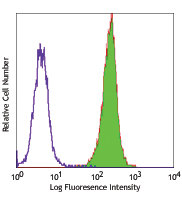
PE anti-human CD326 (EpCAM)

Human colon carcinoma cell line HT29 was stained with CD326 ... -
APC anti-human CD326 (EpCAM)

Human colon carcinoma cell line HT29 stained with 9C4 APC -
Alexa Fluor® 488 anti-human CD326 (EpCAM)

Human colon carcinoma cell line HT29 stained with 9C4 Alexa ... 
Confocal image of human jejunum sample acquired using the IB... 
Confocal image of human jejunum sample acquired using the IB... -
Alexa Fluor® 647 anti-human CD326 (EpCAM)
9C4_Alx647_071007")
Human colon carcinoma cell line (HT29) stainined with 9C4 Al... 
MCF7 breast cancer cell line was stained with 4 µg/mL anti-h... -
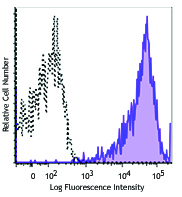
PerCP/Cyanine5.5 anti-human CD326 (EpCAM)

Human colon carcinoma cell line HT-29 was stained with CD326... -
Biotin anti-human CD326 (EpCAM)

Human colon carcinoma cell line (HT29) stained with biotinyl... -
Pacific Blue™ anti-human CD326 (EpCAM)

Human colon carcinoma cell line (HT29) stained with 9C4 Paci... -
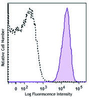
Brilliant Violet 421™ anti-human CD326 (EpCAM)

Human colon carcinoma cell line HT29 was stained with 9C4 Br... -
PE/Cyanine7 anti-human CD326 (EpCAM)

HT-29 cells were stained with CD326 (EpCAM) PE/Cyanine7 (fil... -
Brilliant Violet 605™ anti-human CD326 (EpCAM)

Human colon carcinoma cell line HT29 was stained with CD326 ... -
Brilliant Violet 650™ anti-human CD326 (EpCAM)

Human colon carcinoma cell line HT29 was stained with CD326 ... -
Alexa Fluor® 594 anti-human CD326 (EpCAM)

Human paraffin-embedded placenta tissue slices were prepared... 
Human colorectal adenocarcinoma cell line HT-29 was fixed wi... 
Confocal image of human metastatic lymph node sample acquire... -
Purified anti-human CD326 (EpCAM) (Maxpar® Ready)

Human HT-29 colon carcinoma cells (top) and human Jurkat T c... -
PE/Dazzle™ 594 anti-human CD326 (EpCAM)

Human colon carcinoma cell line HT29 was stained with CD326 ... -
APC/Fire™ 750 anti-human CD326 (EpCAM)

Human colon carcinoma cell line HT29 was stained with CD326 ... -
Brilliant Violet 510™ anti-human CD326 (EpCAM)

Human colon carcinoma cell line HT29 was stained with CD326 ... -
Brilliant Violet 785™ anti-human CD326 (EpCAM)

Human colon carcinoma cell line HT29 was stained with CD326 ... -
Brilliant Violet 711™ anti-human CD326 (Ep-CAM)

Human colon carcinoma cell line HT29 was stained with CD326 ... -
TotalSeq™-A0123 anti-human CD326 (Ep-CAM)
-
APC/Cyanine7 anti-human CD326 (Ep-CAM)

Human colon carcinoma cell line HT29 was stained with CD326 ... -
Alexa Fluor® 700 anti-human CD326 (EpCAM)

Human colon carcinoma cell line HT29 was stained with CD326 ... -
TotalSeq™-C0123 anti-human CD326 (Ep-CAM)
-
TotalSeq™-B0123 anti-human CD326 (Ep-CAM)
-
PE/Cyanine5 anti-human CD326 (Ep-CAM)

Human colon carcinoma cell line, HT29 was stained with CD326... -
TotalSeq™-D0123 anti-human CD326 (Ep-CAM)
-
Spark UV™ 387 anti-human CD326 (Ep-CAM)

Human colon carcinoma cell line HT29 was stained with anti-h... -
Spark Red™ 718 anti-human CD326 (Ep-CAM) (Flexi-Fluor™)


Follow Us