 Login / Register


- Clone
- HI9a (See other available formats)
- Regulatory Status
- RUO
- Workshop
- V P018
- Other Names
- Tetraspanin, MRP-1, DRAP-24
- Isotype
- Mouse IgG1, κ
- Ave. Rating
- Submit a Review
- Product Citations
- publications

-

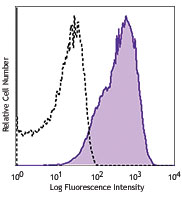
Human peripheral blood platelets stained with HI9a PE
| Cat # | Size | Price | Quantity Check Availability | Save | ||
|---|---|---|---|---|---|---|
| 312105 | 25 tests | £84 | ||||
| 312106 | 100 tests | £184 | ||||
CD9 is a 24 kD type III transmembrane protein also known as tetraspanin, MRP-1 and DRAP-24. It is a member of the tetraspan family (spanning the membrane four times) found on platelets, B cell progenitors, activated lymphocytes, granulocytes, endothelial cells and epithelial cells. CD9 induces adhesion, platelet aggregation, and B cell development. CD9 has been shown to associate with CD63, CD81, CD82, and CD36 and to bind to β1 integrins.
Product DetailsProduct Details
- Verified Reactivity
- Human
- Reported Reactivity
- African Green, Baboon, Cow, Cynomolgus, Dog, Horse, Rabbit, Rhesus, Sheep
- Antibody Type
- Monoclonal
- Host Species
- Mouse
- Formulation
- Phosphate-buffered solution, pH 7.2, containing 0.09% sodium azide and BSA (origin USA)
- Preparation
- The antibody was purified by affinity chromatography, and conjugated with PE under optimal conditions.
- Concentration
- Lot-specific (to obtain lot-specific concentration and expiration, please enter the lot number in our Certificate of Analysis online tool.)
- Storage & Handling
- The antibody solution should be stored undiluted between 2°C and 8°C, and protected from prolonged exposure to light. Do not freeze.
- Application
-
FC - Quality tested
- Recommended Usage
-
Each lot of this antibody is quality control tested by immunofluorescent staining with flow cytometric analysis. For flow cytometric staining, the suggested use of this reagent is 5 µl per million cells in 100 µl staining volume or 5 µl per 100 µl of whole blood.
- Excitation Laser
-
Blue Laser (488 nm)
Green Laser (532 nm)/Yellow-Green Laser (561 nm)
-
Application References
(PubMed link indicates BioLegend citation) -
- Schlossman S, et al. Eds. 1995. Leucocyte Typing V. Oxford University Press. New York.
- Product Citations
-
- RRID
-
AB_2075893 (BioLegend Cat. No. 312105)
AB_2075892 (BioLegend Cat. No. 312106)
Antigen Details
- Structure
- Tetraspan family, type III transmembrane protein, 24 kD
- Distribution
-
Platelets, B cell progenitors, activated lymphocytes, granulocytes, endothelial and epithelial cells
- Function
- Adhesion, platelet activation, B cell development
- Ligand/Receptor
- Associates with CD63, CD81, CD82 and CD36, binds PSG17
- Cell Type
- B cells, Embryonic Stem Cells, Endothelial cells, Epithelial cells, Granulocytes, Lymphocytes, Platelets
- Biology Area
- Immunology, Stem Cells
- Molecular Family
- CD Molecules
- Antigen References
-
1. Miao WM, et al. 2001. Blood 97:1689.
2. Ellerman DA, et al. 2003. Mol Biol Cell. 14:5098.
3. Schlossman S, et al. Eds. 1995. Leucocyte Typing V. Oxford University Press. New York. - Gene ID
- 928 View all products for this Gene ID
- UniProt
- View information about CD9 on UniProt.org
Related Pages & Pathways
Pathways


Related FAQs
- What type of PE do you use in your conjugates?
- We use R-PE in our conjugates.
Other Formats
View All CD9 Reagents Request Custom Conjugation| Description | Clone | Applications |
|---|---|---|
| Purified anti-human CD9 | HI9a | FC,ICC |
| FITC anti-human CD9 | HI9a | FC |
| PE anti-human CD9 | HI9a | FC |
| APC anti-human CD9 | HI9a | FC |
| PerCP/Cyanine5.5 anti-human CD9 | HI9a | FC |
| APC/Fire™ 750 anti-human CD9 | HI9a | FC |
| Biotin anti-human CD9 | HI9a | FC |
| PE/Cyanine7 anti-human CD9 | HI9a | FC |
| PE/Dazzle™ 594 anti-human CD9 | HI9a | FC |
| TotalSeq™-A0579 anti-human CD9 | HI9a | PG |
| TotalSeq™-C0579 anti-human CD9 | HI9a | PG |
| TotalSeq™-B0579 anti-human CD9 Antibody | HI9a | PG |
| TotalSeq™-D0579 anti-human CD9 | HI9a | PG |
| APC/Fire™ 750 anti-human CD9 | HI9a | FC |
| FITC anti-human CD9 | HI9a | FC |
| APC anti-human CD9 | HI9a | FC |
| Spark Violet™ 423 anti-human CD9 | HI9a | FC |
Customers Also Purchased
Compare Data Across All Formats
This data display is provided for general comparisons between formats.
Your actual data may vary due to variations in samples, target cells, instruments and their settings, staining conditions, and other factors.
If you need assistance with selecting the best format contact our expert technical support team.
-
Purified anti-human CD9

Human platelets stained with purified HI9a, then detected wi... 
BT474 breast cancer cell line was stained with anti-human CD... -
FITC anti-human CD9

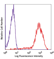
Human peripheral blood platelets stained with HI9a FITC -
PE anti-human CD9

Human peripheral blood platelets stained with HI9a PE -
APC anti-human CD9

Human platelets were stained with CD9 (clone HI9a) APC (fill... -
PerCP/Cyanine5.5 anti-human CD9

Human platelets were stained with CD9 (clone HI9a) PerCP/Cya... -
APC/Fire™ 750 anti-human CD9

Human platelets were stained with CD9 (clone HI9a) APC/Fire™... -
Biotin anti-human CD9

Human platelets were stained with biotinylated CD9 (clone HI... -
PE/Cyanine7 anti-human CD9

Human platelets were stained with CD9 (clone HI9a) PE/Cyanin... -
PE/Dazzle™ 594 anti-human CD9

Human platelets were stained with CD9 (clone HI9a) PE/Dazzle... -
TotalSeq™-A0579 anti-human CD9
-
TotalSeq™-C0579 anti-human CD9
-
TotalSeq™-B0579 anti-human CD9 Antibody
-
TotalSeq™-D0579 anti-human CD9
-
APC/Fire™ 750 anti-human CD9

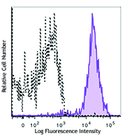
Typical results from human peripheral blood platelets staine... -
FITC anti-human CD9

Typical results from human peripheral blood platelets staine... -
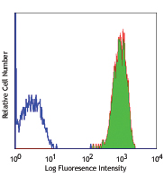
APC anti-human CD9

Typical results from human peripheral blood platelets staine... -
Spark Violet™ 423 anti-human CD9

Human platelets were stained with anti-human CD9 (clone HI9a...


Follow Us